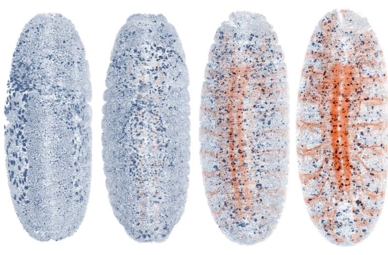
Light-sheet microscopy is one of the most powerful method for imaging the development and function of whole living organisms. However, achieving high-resolution images with these microscopes requires manual adjustments during imaging. Researchers of the Max Planck Institute of Molecular Cell Biology and Genetics in Dresden together with colleagues at Janelia Research Campus (HHMI) have developed a new kind of light-sheet microscope that can ‘drive’ itself automatically by adapting to the challenging and dynamic optical conditions of large living specimens. This new smart microscope combines a novel hardware design and a smart ‘AutoPilot’ system that can analyze images and automatically adjust and optimize the microscope. This framework enables for the first time long-term adaptive imaging of entire developing embryos and improves the resolution of light-sheet microscopes up to five-fold.
Light sheet microscopy is a novel microscopy technique developed in the last ten years that is uniquely suited to image large living organisms. In a light-sheet microscope, a laser light sheet illuminates the sample perpendicularly to the observation along a thin plane within the sample. Out-of-focus and scattered light from other planes—which often impair image quality—is largely avoided because only the observed plane is illuminated.
The long-standing goal of microscopy is to achieve ever-sharper images deep inside of living samples. For light-sheet microscopes this requires to perfectly maintain the careful alignments between imaging and light-sheet illumination planes. Mismatches between these planes arise from the optical variability of living tissues across different locations and over time. Tackling this challenge is essential to acquire the high-resolution images necessary to decipher the biology behind organism development and morphogenesis. “So far, researchers had to sit at their microscope and tweak things manually—our system puts an end to this: it is like a self-driving car: it functions autonomously”, says Loïc Royer, first author of the study. This smart autonomous microscope can in real-time analyze and optimize the spatial relationship between light-sheets and detection planes across the specimen volume.
