Toggle light / dark theme

The researchers turned to a group of molecules called acylcarnitines, which are associated with declining cognition and breaking down or metabolizing fats and proteins for energy. To test if high acylcarnitine levels in the blood could predict who’s at risk of developing Alzheimer’s, the researchers used data from a large-scale study called the Alzheimer’s Disease Neuroimaging Initiative.

“It was fascinating,” the author said. “Dividing research participants into groups based on their specific acylcarnitine levels highlighted people with more severe Alzheimer’s disease and others with fewer symptoms.” This led the researchers to define a bioenergetic clock based on acylcarnitines—how old a person’s metabolism acts, compared to actual age. Higher bioenergetic age is linked to higher acylcarnitine levels, worsened Alzheimer’s pathology, cognitive decline and brain atrophy.

The researchers also quantified cognitive decline using a common test called the mini-mental state examination, on which a score below 24 out of 30 points indicates impairment. They found that people with low acylcarnitine levels to begin with declined more slowly, losing about 0.5 points less per year than people with high acylcarnitine levels. The benefit is on par with the Alzheimer’s drug lecanemab.

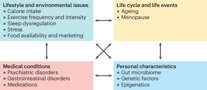
To some degree, a person’s bioenergetic clock ticks forward at a rate determined by their genetics, but having a healthy lifestyle—for example, eating a plant-based diet and exercising —can help keep acylcarnitine levels low, which means a younger bioenergetic age, the author explained.

They went on to identify a subgroup of participants, about 30% of the Alzheimer’s Disease Neuroimaging Initiative, with older bioenergetic age but favorable genetic background. These individuals may benefit more from early lifestyle interventions designed to decrease their bioenergetic age and potentially delay or prevent the onset of Alzheimer’s.

Moving forward, the senior author hopes to home in on the lifestyle interventions most effective for lowering bioenergetic age. For example, eating a low-carb diet may help maintain metabolic health, but just how low would carbohydrate consumption have to be for a person to see benefits?


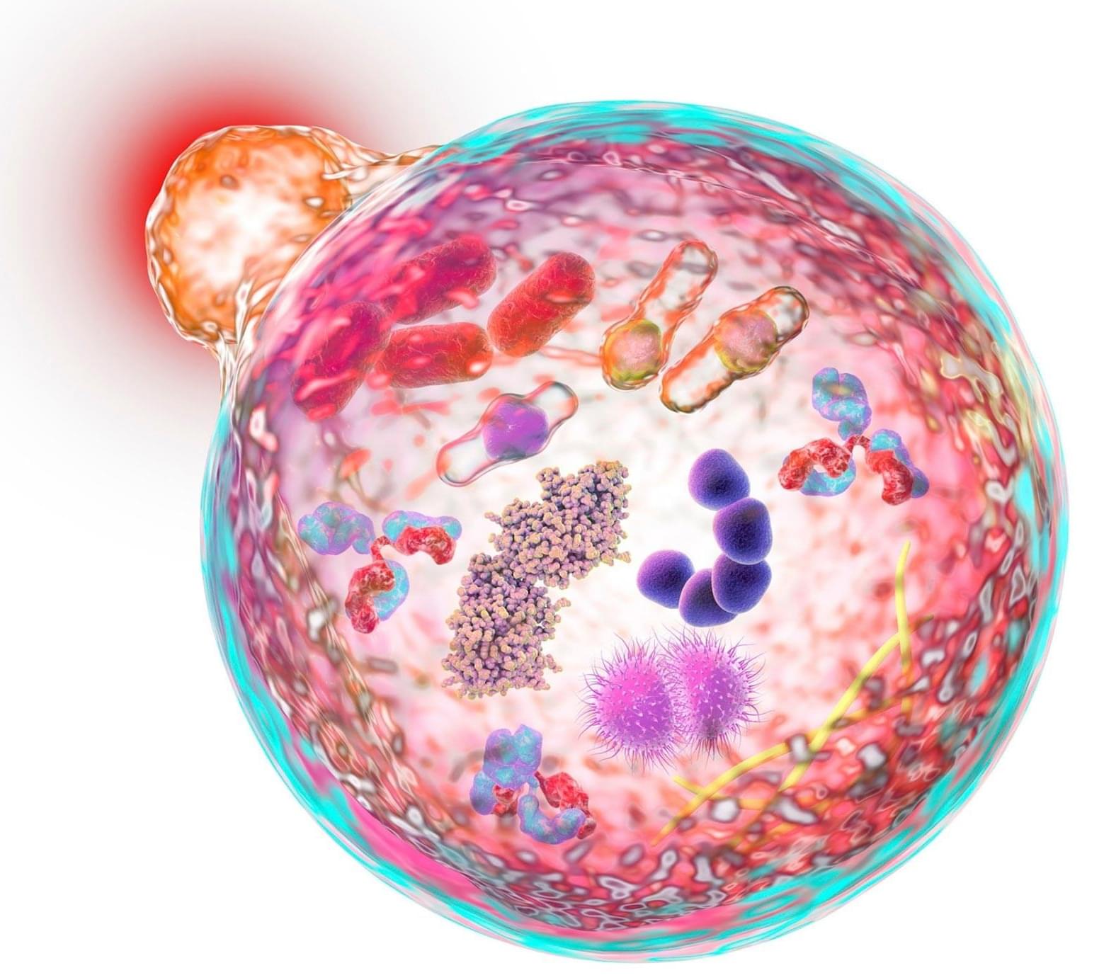
A drug-resistant type of bacteria that has adapted to health care settings evolved in the past several years to weaponize an antimicrobial genetic tool, eliminating its cousins and replacing them as the dominant strain. University of Pittsburgh School of Medicine scientists made the discovery when combing through local hospital data—and then confirmed that it was a global phenomenon.

The finding, published in Nature Microbiology, may be the impetus for new approaches in developing therapeutics against some of the world’s deadliest . It also validates a new use for a system developed at Pitt and UPMC that couples genomic sequencing with computer algorithms to rapidly detect infectious disease outbreaks.

“Our lab has a front row seat to the parade of pathogens that move through the ,” said senior author Daria Van Tyne, Ph.D., associate professor of medicine in Pitt’s Division of Infectious Diseases. “And when we took a step back and zoomed out, it quickly became apparent that big changes were afoot with one of the world’s more difficult-to-treat bacteria.”

Additional experiments revealed that mice given access to an exercise wheel or treated with antidepressants also exhibited increased CB1 receptor levels in astrocytes. Furthermore, analysis of human brain tissue from the Douglas-Bell Canada Brain Bank indicated that individuals with major depression had lower astrocytic CB1 receptor levels compared to those without depression or those who had received antidepressant treatment.

Implications for mental health interventions

These findings raise the possibility of developing treatments that selectively activate CB1 receptors in astrocytes to mitigate anxiety and depression. However, the challenge remains in limiting activation to astrocytes, as prolonged CB1 receptor stimulation in neurons can lead to side effects affecting alertness, anxiety and appetite. Until targeted pharmacological interventions become available, engaging in physical activity may help protect against stress-related mental health conditions by enhancing CB1 receptor expression.

A new study by UCLA Health has discovered what researchers say is the first drug to fully reproduce the effects of physical stroke rehabilitation in model mice, following from human studies.

The findings, published in Nature Communications, tested two candidate drugs derived from their studies on the mechanism of the brain effects of rehabilitation, of which one resulted in significant recovery in movement control after stroke in the mouse model.

Stroke is the leading cause of adult disability because most patients do not fully recover from the effects of stroke. There are no drugs in the field of stroke recovery, requiring stroke patients to undergo physical rehabilitation which has shown to be only modestly effective.

A study from Tübingen University and the German Center for Diabetes Research reveals that the brain plays a crucial role in obesity and type 2 diabetes development. It shows that even a brief period of consuming high-calorie processed foods can significantly alter brain insulin sensitivity, a key factor in weight gain and metabolic disorders. The research demonstrated that insulin’s appetite-suppressing effect in the brain diminishes after a short-term high-calorie diet, leading to insulin resistance. These effects were observed in healthy participants, suggesting that dietary habits could influence brain function before any significant weight gain occurs. Further research is needed to understand the brain’s role in these conditions.


The number of obese persons has grown significantly in recent decades, which presents significant difficulties for those who are impacted, healthcare systems, and those who provide treatment. The hormone insulin plays a key role in the development of obesity. Up until recently, there have been numerous signs indicating insulin causes neurodegenerative and metabolic disorders, especially in the brain. A recent study by the University Hospital of Tübingen, the German Center for Diabetes Research (DZD), and Helmholtz Munich offers intriguing new insights into the origins of type 2 diabetes and obesity as well as the brain’s function as a critical control center.

Obesity has only been officially recognized as a disease in Germany since 2020, despite the fact that it has long been known to cause a number of illnesses, including diabetes, heart attacks, and even cancer. The World Health Organization has already declared obesity to be an epidemic, affecting over one billion individuals globally and almost 16 million in Germany alone. A body mass index of 30 or more is considered obese, and a poor diet and insufficient exercise are frequently cited as the causes of this chronic illness. However, the mechanisms in the body that lead to obesity and cause the disease are more complex.

Obesity and the role of insulin in the brain

Unhealthy body fat distribution and chronic weight gain are linked to the brain’s sensitivity to insulin. What specific functions does insulin perform in the brain, and how does it affect individuals of normal weight? In their study, Prof. Dr. Stephanie Kullmann and her colleagues at the Tübingen University Hospital for Diabetology, Endocrinology, and Nephrology found the answer to this query. “Our findings demonstrate for the first time that even a brief consumption of highly processed, unhealthy foods (such as chocolate bars and potato chips) causes a significant alteration in the brain of healthy individuals, which may be the initial cause of obesity and type 2 diabetes,” says Prof. Kullmann, the study’s leader. In a healthy state, insulin has an appetite-suppressing effect in the brain. However, in people with obesity in particular, insulin no longer regulates eating behavior properly, resulting in insulin resistance.

How does the armored tiling on shark and ray cartilage maintain a continuous covering as the animals’ skeletons expand during growth?

This is a question that has perplexed Professor Mason Dean, a in the Department of Infectious Diseases and Public Health at City University of Hong Kong (CityUHK) since he was in graduate school.

An expert in , structure and function in vertebrate animals, but with a particular focus on (and affection for) sharks and rays, Professor Dean says he was curious about how nature keeps complex surfaces covered while organs and animals are growing, and their surfaces are changing.