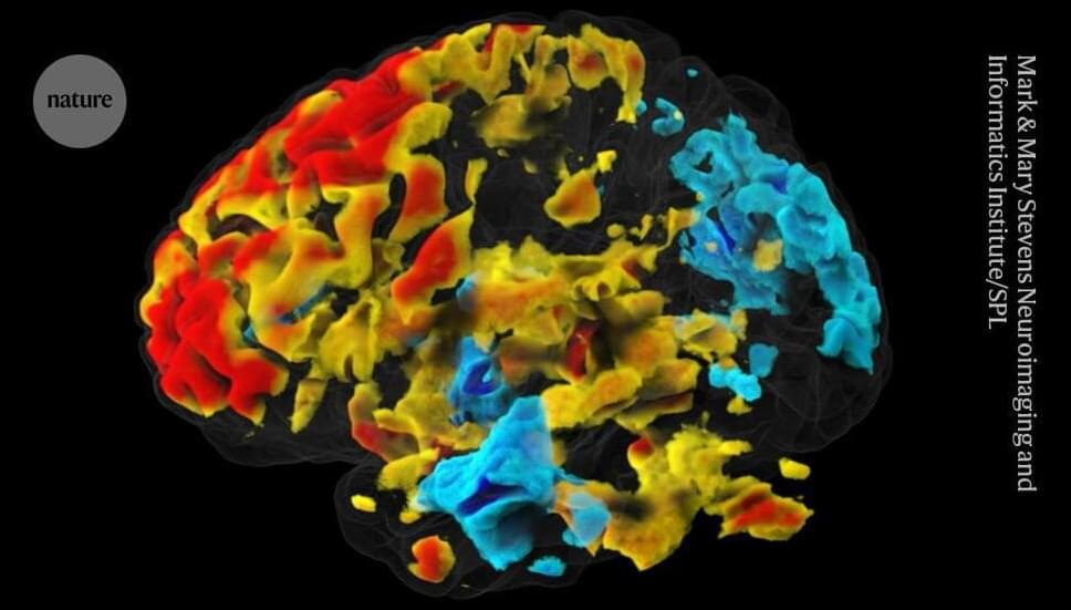
In conventional functional MRI (fMRI), researchers monitor changes in blood flow to different brain regions to estimate activity. But this response lags by at least one second behind the activity of neurons, which send messages in milliseconds.
Park and his co-authors said that DIANA could measure neuronal activity directly, which is an “extraordinary claim”, says Ben Inglis, a physicist at the University of California, Berkeley.
The DIANA technique works by applying minor electric shocks every 200 milliseconds to an anaesthetized animal. Between shocks, an MRI scanner collects data from one tiny piece of the brain every 5 milliseconds. After the next shock, another spot is scanned. The software stitches together data from all the spots, to visualize changes in an entire slice of brain over a 200-millisecond period. The process is similar to filming an action pixel by pixel, where the action would need to be repeated to record every pixel, and those recordings stitched together, to create a full video.
