Toggle light / dark theme

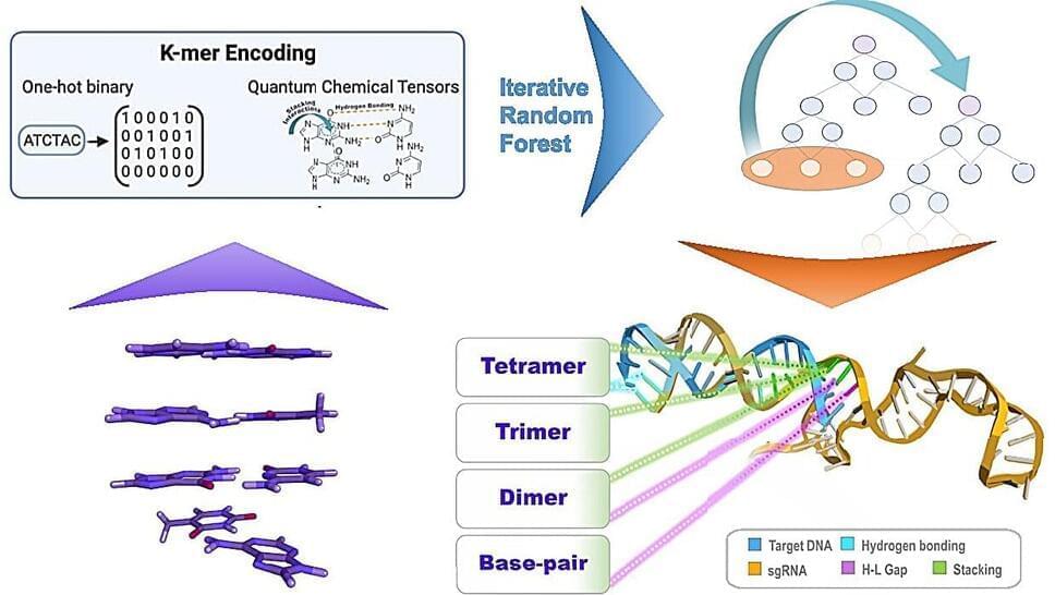
A new technique has been added to the CRISPR gene-editing toolbox. Known as PASTE, the system uses virus enzymes to “drag-and-drop” large sections of DNA into a genome, which could help treat a range of genetic diseases.

The CRISPR system originated in bacteria, which used it as a defense mechanism against viruses that prey on them. Essentially, if a bacterium survived a viral infection, it would use CRISPR enzymes to snip out a small segment of the virus DNA, and use that to remind itself how to fight off future infections of that virus.

Over the past few decades, scientists adapted this system into a powerful tool for genetic engineering. The CRISPR system consists of an enzyme, usually one called Cas9, which cuts DNA, and a short RNA sequence that guides the system to make this cut in the right section of the genome. This can be used to snip out problematic genes, such as those that cause disease, and can substitute them with other, more beneficial genes. The problem is that this process involves breaking both strands of DNA, which can be difficult for the cell to patch back up as intended, leading to unintended alterations and higher risks of cancer in edited cells.

Unnecessary playing with nature.


In January, Bennett’s doctors offered him the chance to receive a heart from a pig. He took it. “I know it’s a shot in the dark, but it’s my last choice,” he said in a press release from the University of Maryland Medical Center in Baltimore, where he was being treated. On 7 January, doctors transplanted the heart, which had been genetically modified so that the human body would tolerate it.

Bennett survived for eight weeks with his new heart before his body shut down. After his death, the research team learnt that the transplanted organ was infected with a pig herpesvirus that had not been detected by tests1.

But even a few weeks is a long time for an animal organ placed in a human, known as a xenotransplant. Given that the human immune system begins attacking non-genetically modified pig organs in minutes, other xenotransplantation researchers are impressed with the experiment. “It’s actually beyond my expectation that the patient lived up to two months,” says Luhan Yang, a bioengineer and chief executive of Qihan Biotech in Hangzhou, China. “I think it’s a victory for the field.”

Evidence-Based And Actionable Health, Wellness And Longevity Solutions — Dr. Renee DeHaan, Ph.D. — VP, Science & AI, InsideTracker


Dr. Renée Deehan, Ph.D. is the VP of Science & Artificial Intelligence at InsideTracker (https://www.insidetracker.com/), and leads a science team that builds and mines the world’s largest data set of blood, DNA, fitness tracking and phenotypic data from healthy people, creating evidence-based solutions that are simple, clear, and actionable.

Dr. Deehan has spent her career working in the precision medicine and personalized nutrition domains, previously serving as the VP of Computational Biology & Translational Informatics at QuartzBio and as the VP of Biology and Bioinformatics at PatientsLikeMe, the world’s largest integrated community, health management, and real-world data platform.

Circa 2021:3


Surgical management of breast cancer often results in the absence of the breast. However, existing breast reconstruction methods may not meet the need for a replacement tissue. Tissue engineering with the use of emerging materials offers the promise of generating appropriate replacements. Three-dimensional (3D) printing technology has seen a significantly increased interest and application in medically-related fields in the recent years. This has been especially true in complex medical situations particularly when abnormal or complicated anatomical surgical considerations or precise reconstructive procedures are contemplated. In addition, 3D bio-printing which combines cells with bio-material scaffolds offers an exciting technology with significant applications in the field of tissue engineering. The purpose of this manuscript was to review a number of studies in which 3D printing technology has been used in breast reconstructive surgical procedures, and future directions and applications of 3D bio-printing.

Breast cancer is the most common cancer diagnosed among US women and is second only to lung cancer as a cause of cancer death among women as of 2019. Because ~268,600 (almost six times than DCIS) new cases prove to be an invasive type of breast cancer (1), many women had to choose the removal of the breast, with immediate consideration for a replacement tissue. Although this was satisfactory in many patients, either saline or gel-filled breast implants (2) do carry real risks of complications such as infection, capsular contracture, implant dislocation, or deformities (3, 4). The option of autologous reconstruction can be more texturally natural aesthetically, but it requires a more complex procedure, significant time and expense, and possible muscle weakness or hernia formation at the tissue donor site (5). Tissue engineering intends to address these limitations by combining the 3D printing technology with synthetic or natural structural elements.

Three-dimensional (3D) printing, also known as computer-aided manufacturing (CAM), was based on digital model files using metal powder or plastic and other adhesive materials to construct objects with a computer guided precision, printing layer upon layer. Simplistically, it uses a computer aided design (CAD) program to convert the virtual model of an object into a printable object using an STL (Standard Tessellation Language or STereoLithography) file. The object then gradually and precisely takes shape as each thin layer is added according to the design file, and composed of the desired material for that object in the form of “ink” using the 3D printer. Not only in cases of intraoperative 3D printed models serving as templates, but this technology has extended to implanted scaffolds that have been used to correct defect-specific sites, clearly enhancing patient treatment (6, 7).

Researchers believe that synthetic muscle fibers could be used in a wide variety of sustainable and environmentally friendly industrial applications, including textiles, biomedicine, and tissue engineering. In a world where it takes up to 1,800 gallons of water to produce a single pair of jeans, the development of such environmentally safe processes that provide high-strength materials for industrial applications is a great step forward.


How strong can a muscle ever get? Can it have more endurance than metal? Can it be sturdier than Kevlar? While you might be inclined to answer the above in the negative, please pause, for scientists have succeeded in developing synthetic muscle that’s stronger than Kevlar. How about that for a flex?

The science and other stuff to know

The breakthrough was achieved by researchers at the McKelvey School of Engineering at Washington University, and the findings were reported in a study published in Nature Communications in August.

High-Risk, High-Payoff Bio-Research For National Security Challenges — Dr. David A. Markowitz, Ph.D., IARPA


Dr. David A. Markowitz, Ph.D. (https://www.markowitz.bio/) is a Program Manager at the Intelligence Advanced Research Projects Activity (IARPA — https://www.iarpa.gov/) which is an organization that invests in high-risk, high-payoff research programs to tackle some of the most difficult challenges of the agencies and disciplines in the U.S. Intelligence Community (IC).

IARPA’s mission is to push the boundaries of science to develop solutions that empower the U.S. IC to do its work better and more efficiently for national security. IARPA does not have an operational mission and does not deploy technologies directly to the field, but instead, they facilitate the transition of research results to IC customers for operational application.

b Department of Polymer Science and Engineering and Key Laboratory of High Performance Polymer Materials and Technology of MOE, School of Chemistry and Chemical Engineering, Nanjing University, Nanjing 210,023, China.

c Institute of Chemical and Bioengineering, ETH Zurich, Vladimir Prelog Weg 1, 8093 Zurich, Switzerland.

Received 21st February 2019, Accepted 17th April 2019.

The fourth discussion of the NEW NOW program, “Transhumanism: Beyond the Human Frontier?”, took place on December 16.

Together with our guest experts, we tried to identify the latest technology that has either already become a reality or is currently in development, focusing on the ethical aspects of the consequences that ensue. We reflected on the question of whether the realization of transhumanist ideas is likely to entail a radical change in the ways people relate to one another. How far are we prepared to go in changing our bodies in order to attain these enhanced capacities? We will attempt to identify the “human frontier”, beyond which the era of posthumanism awaits.

Speakers:

James “J.” Hughes Ph.D. is a bioethicist and sociologist who serves as the Associate Provost for the University of Massachusetts Boston (UMB), and as Senior Research Fellow at UMB’s Center for Applied Ethics. He holds a doctorate in Sociology from the University of Chicago where he taught bioethics at the MacLean Center for Clinical Medical Ethics. Since then Dr. Hughes has taught health policy, bioethics, medical sociology and research methods at Northwestern University, the University of Connecticut, and Trinity College.

A team of researchers from Purdue University claim to have discovered the “chemistry behind the origin of life” on Earth in simple droplets of water, and they’re using strikingly strong language to celebrate the findings.

Graham Cooks, chemistry professor at Purdue and lead author of a new paper published in the journal Proceedings of the National Academy of Sciences, called it a “dramatic discovery” and the “secret ingredient for building life” in a statement.

“This is essentially the chemistry behind the origin of life,” he added. “This is the first demonstration that primordial molecules, simple amino acids, spontaneously form peptides, the building blocks of life, in droplets of pure water.”