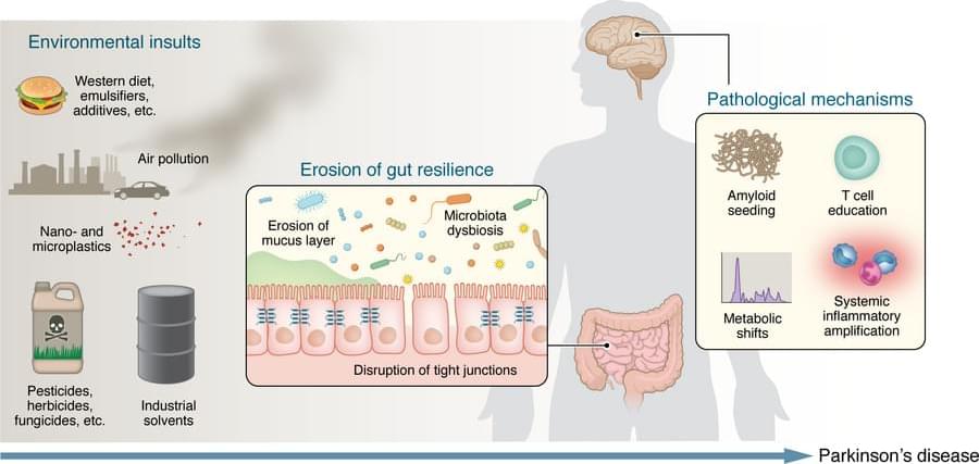
Toggle light / dark theme

Applying tissue maturation techniques to engineered cartilage grafts produces more functionally faithful grafts and leads to superior clinical outcomes in patients with knee cartilage injuries, shows a new multicenter clinical trial.

📄


Engineered hyaline-like cartilage tissues are superior to immature cell-based grafts for the therapy of cartilage defects in the human knee.

Scientists have now cracked this secret using computational simulations and lab experiments, paving the way for bioengineered silk with game-changing applications, from medical sutures to ultra-strong body armor.

Spiders Strengthen Their Silk with Stretching

When spiders spin their webs, they use their hind legs to pull silk from their spinnerets. This pulling action does more than just release the silk—it strengthens the fibers, making the web more durable.

Half a million people in the UK with dangerously high blood pressure – a “silent killer” that causes tens of thousands of deaths a year – could be cured by a new treatment.

Doctors have developed a technique to burn away nodules that lead to a large amount of salt building up in the body, which increases the risk of a stroke or heart attack.

The breakthrough could mean people with primary aldosteronism – which causes one in 20 cases of high blood pressure – no longer have to have surgery or spend their lives taking the drug spironolactone to lower their risk of a stroke or heart attack.

We finally have a more natural method to kill cancer.

A study from Cold Spring Harbor Laboratory suggests that a vitamin K precursor, menadione, may offer a highly targeted way to kill prostate cancer cells.

Unlike traditional treatments that push cancer into dormancy, menadione acts as a pro-oxidant, disrupting a key lipid called PIP. This lipid helps cells manage waste, and without it, cancer cells become overwhelmed and ultimately burst.

The study, published in Science, demonstrated significant tumor suppression in both mice and human cancer cells. Researchers believe this method could offer a safer and more definitive resolution for prostate cancer while minimizing the risk of resistance.

Beyond cancer, menadione also shows promise in treating X-linked myotubular myopathy, a severe genetic muscle disorder. Importantly, menadione’s safety profile appears favorable, as it is commonly used in animal feed to support vitamin K production.

The findings suggest that menadione could be especially beneficial for prostate cancer patients under active surveillance, potentially delaying or even preventing progression.

With low side effects and a highly selective approach, this research offers new hope for effective, minimally invasive cancer treatment options.

To mimic the conditions of the human brain, the researchers opted not to use a mouse model for MS, instead advancing a model that uses the marmoset, a nonhuman primate. Compared to mouse brains, marmoset and human brains have a higher ratio of white matter (the “wires” of the brain) to gray matter (neuronal cell bodies). The marmoset model creates multiple lesions that closely resemble those seen in human MS and that can be tracked in real time using MRI imaging. Because these lesions can be induced experimentally, the model offers a look at the earliest stages of inflammation and immune responses that lead to MS-like demyelination.

One key player identified was a specific type of astrocyte, one of the support cell types in the brain, that turns on a gene called SERPINE1 or plasminogen activator inhibitor-1 (PAI1). They found SERPINE1-expressing astrocytes in vulnerable brain borders before visible damage occurs, clustering near blood vessels and the fluid-filled ventricles of the brain and signaling future areas of lesion development. These astrocytes also appeared to influence the behavior of other cells near the lesion area, including the ability of immune cells to enter the brain and contribute to inflammation, as well as the precursor cells involved in myelin repair.

Given that SERPINE1-expressing astrocytes accumulated at the edges of growing lesions, where damage happens but healing also begins, their potential dual role in coordinating signals that could lead to either tissue repair or further damage was an unexpected wrinkle that will require further study. It’s possible that the earliest responses could be a part of a protective mechanism that becomes overwhelmed as the injury progresses. It’s also possible that the same mechanism could itself become disease-causing.


Using an animal model of multiple sclerosis (MS), researchers have created a four-dimensional brain map that reveals how lesions similar to those seen in human MS form. These findings, published in Science, provide a window into the early disease state and could help identify potential targets for MS treatments and brain tissue repair.

The researchers combined repeated MRI imaging with brain-tissue analysis, including gene expression, to track the onset and development of MS-like lesions. They uncovered a new MRI signature that can help detect brain regions at risk for damage weeks before any visible lesions occur. They also identified “microenvironments” within affected brain tissue based on observed patterns of neural function, inflammation, immune and support cell responses, gene expression, and levels of damage and repair.

“Identifying the early events that occur after inflammation and teasing apart which are reparative versus which are damaging, can potentially help us identify MS disease activity sooner and develop treatments to slow or stop its progression,” said the author.

The discovery of a mini aurora above a light-emitting polymer material reveals an electron-ejection process that might be useful in field-emission displays and material fabrication.

Auroras occur in the night sky when charged solar-wind particles, such as protons and electrons, are deflected by Earth’s magnetic field and interact with molecules in the atmosphere. Researchers have now found an aurora-like emission coming from a light-emitting polymer [1]. The surprising display consisted of flashes of green light above the polymer surface. The researchers explained the emission as the result of electrons being ejected from the polymer and interacting with a vapor of organic molecules. The discovery suggests that these polymers might be useful as electron emitters for applications such as spectroscopy, medical technology, and lithography.

Jun Gao from Queen’s University in Canada is amazed by auroras, and he’s even gone out on cold nights to look for them. But he was not prepared for the aurora that showed up in his lab two years ago. He and his student at the time, Dongze Wang, were testing failure modes for polymer light-emitting electrochemical cells, or PLECs, used in light sources and display devices. These cells are organic semiconductors that are electrochemically doped on one side to have excess electrons (making an n-type semiconductor) and on the other side to have electron deficiencies, or holes (making a p-type semiconductor). Electrons crossing the p – n boundary can fill holes and produce red light.

A team of cognitive neuroscientists and acoustic engineers at Adam Mickiewicz University, in Poland, has found no evidence that wind turbine noise causes mental impairment. In their study, published in the journal Humanities and Social Sciences Communication, the group conducted experiments exposing human volunteers to various noises and measured a range of impacts.

Over the past several years, several groups and individuals around the world, most particularly in the U.S., have conceived of the idea of something called “wind turbine syndrome”—a theory that suggests noise from windmills can cause , or other health problems such as cancer. To date, such claims have not been backed up by research or any other type of proof. In this new effort, the research team in Poland sought to find out if there is any merit to the theory.

The researchers recruited 45 students at a local university who listened to various noises while wearing devices that measured their brainwaves. The researchers intentionally chose young volunteers because prior research has shown they are more sensitive to noise than .

The brain has higher concentrations of plastic particles compared to other organs, with increased levels found in dementia patients.

In a comprehensive commentary published in Brain Medicine, researchers highlight alarming new evidence of microplastic accumulation in human brain tissue, offering critical insights into potential health implications and prevention strategies. This commentary examines findings from a groundbreaking Nature Medicine article by Nihart et al. (2025) on the bioaccumulation of microplastics in the brains of deceased individuals.

The research reveals that human brains contain approximately a spoonful of microplastics and nanoplastics (MNPs), with levels three to five times higher in individuals with documented dementia diagnoses. Even more concerning, brain tissue exhibited MNP concentrations seven to thirty times higher than those found in other organs, such as the liver or kidneys.

The brain’s ability to process information is known to be supported by intricate connections between different neuron populations. A key objective of neuroscience research has been to delineate the processes via which these connections influence information processing.

Researchers at the University of Padova, the Max Planck Institute for the Physics of Complex Systems and École Polytechnique FĂ©dĂ©rale de Lausanne recently carried out a study aimed at better understanding the contribution of excitatory and inhibitory neuron populations to the brain’s encoding of information. Their findings, published in Physical Review Letters, show that is maximized when the activity of excitatory and inhibitory neurons is balanced.

“Our research was inspired by a fundamental question in neuroscience: how does the structure of the brain shape its ability to process information?” Giacomo Barzon, co-author of the paper, told Medical Xpress. “The brain continuously receives and integrates sensory inputs, and neurons do not act in isolation—they are part of complex, recurrent networks. One particularly intriguing feature of these networks is the balance between the activity of excitatory and inhibitory neurons, which has been observed across different brain regions.”