Toggle light / dark theme

Reference Handbook of Gynecologic Pelvic MRI

Accurate interpretation of gynecologic pelvic MRI depends on tailored protocols, thorough anatomic knowledge, and a systematic lesion-based approach. This review outlines classification by lesion origin and tissue composition.


MRI is used routinely in patients with gynecologic conditions due to its superior soft-tissue contrast and multiplanar capability. It can provide a detailed assessment of pelvic anatomy, lesion origin, tissue characteristics, and disease extent. Its indications include (a) characterizing US-indeterminate adnexal masses, (b) evaluating the cause of pelvic pain and abnormal uterine bleeding, © mapping symptomatic uterine leiomyomas and differentiating them from uterine sarcomas, (d) staging gynecologic cancers, and (e) addressing complex congenital müllerian duct anomalies. Additional indications include evaluating for pelvic floor dysfunction, fistulas, or placental abnormalities.

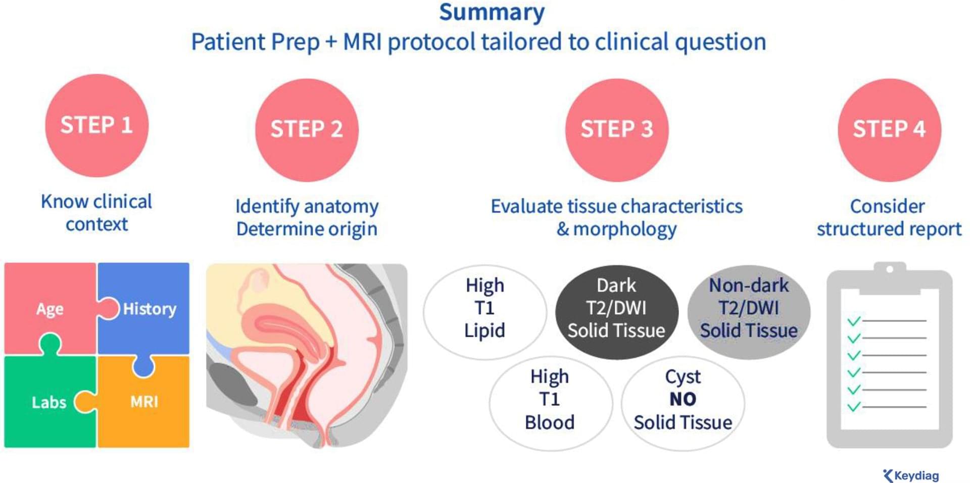
This article aims to enhance the interpretation of female pelvic MRI for gynecologic conditions. After outlining patient preparation, tailored MRI protocols, and the normal female pelvic anatomy, a step-by-step approach to interpretation is presented, focusing on evaluating lesion origin, lesion tissue composition, and solid tissue morphology (Fig 1). The review also emphasizes the importance of incorporating the patient’s clinical history to narrow the differential diagnosis and the role of disease-specific structured reporting in improving communication (Fig 1).

Does suppression of HIV replication protect against severe M

tuberculosis infection?

Philana Ling Lin & team report on SIV/ART/TB models, finding antiretroviral treatment reduces pulmonary TB pathology, yet does not prevent extrapulmonary spread of TB.


1Department of Pediatrics, Children’s Hospital of Pittsburgh of the University of Pittsburgh Medical Center, Pittsburgh, Pennsylvania, USA.

2Center for Vaccine Research.

3Department of Microbiology and Molecular Genetics, and.

4Division of Laboratory Animal Research, University of Pittsburgh School of Medicine, Pittsburgh, Pennsylvania, USA.

PDE5A+ cancer-associated fibroblasts enhance immune suppression in gastric cancer

GUTImage from the paper by Wang et al entitled.

via.

https://bit.ly/4ae9wtW

GastricCancer.


Background Gastric cancer (GC) ranks among the most prevalent lethal tumours globally. Cancer-associated fibroblasts (CAFs) are pivotal in creating an immunosuppressive tumour microenvironment (TME) in GC.

Objective Identifying a critical subpopulation of CAFs in promoting an immunosuppressive TME and enabling immune evasion, which may influence therapeutic effectiveness of immune checkpoint inhibitors (ICIs) for GC.

Standardization Versus Adaptability: Where Is the Sweet Spot?

New in practicalRO.


Standardization in clinical workflows is widely recognized as a driver of safety, efficiency, and consistency. The challenge for modern practice is determining the appropriate degree and rigidity of standardization, especially as automation and adaptive technologies reshape workflows.

Where Biology Meets Resonance: Light, Vibration, and Living Order

When we think about biology, we usually picture chemistry: molecules bumping into each other, enzymes reacting, and signals spreading by diffusion. That picture is real—but it may be incomplete. In my recent paper in Harmonic Science Perspectives (Vol 1, Issue 1), I propose a complementary layer of cellular organization: a fast, coordination-capable “resonance network” that uses three interchangeable carriers of energy and information.

IntroductionA simple picture: three messengers that can translate into one anotherWhere this shows up in the body: mitochondria and microtubules as a coupled networkWhy interconversion matters: translation is the key featureResonant synchronization: a possible mechanism for cellular timingTherapeutic implications: why light and sound therapies might work better togetherA note on what’s established vs what’s proposedConclusion: a new lens on living organization

Those three carriers are light (photons), vibration/sound-like mechanical waves (phonons), and mobile electronic excitations in biomolecules (excitons). The central idea is simple to state even if the details are deep: living systems may continuously convert energy back and forth between these three modes to synchronize activity across space and time inside the cell—and potentially across tissues.

Mechanisms and Regulation of Cellular Senescence

Cellular senescence is generally an irreversible proliferative arrest in damaged normal cells that have exited the cell cycle. These cells display high metabolic activities [1], remain viable, and actively suppress apoptosis [2, 3]. Senescent cells present unique morphological and molecular characteristics and functions that distinguish them from other nondividing cell populations, such as quiescent cells and terminally differentiated cells [4, 5, 6]. The hallmarks of cellular senescence include: prolonged cell cycle arrest, transcriptional changes, acquisition of a bioactive secretome, known as the senescence-associated secretory phenotype (SASP), macromolecular damage, and deregulated metabolism [7].

Replicative senescence was the first cellular senescence subtype to be described [8]. It is induced after serial propagation of normal human cells in culture and is caused by telomere erosion and the consequent increase in DNA lesions [9, 10,11,12]. The limited lifespan of most (perhaps all) cultured primary cells is influenced by the species and tissue type from which they were derived. Senescence can also be triggered by many other intrinsic and extrinsic factors, particularly, replicative stress, oxidative damage, metabolism dysfunctions, cytokines, oncogene activation, and chemotherapy agents. All these factors can induce DNA damage and senescence in normal and cancer cells (in some contexts) [6]. Cellular senescence occurs not only in vitro (i.e., cell culture models), but also in various tissues in vivo [13,14,15,16].

Senescence is an important contributor to cancer and aging, two processes characterized by a time-dependent accumulation of cell damage and dysfunction. Senescence markers are detected in premalignant tumor lesions but not at later stages of tumor development [17,18,19]. The proliferative arrest imposed by cellular senescence represents an early barrier against cancer initiation by preventing the propagation of damaged DNA to the next generation of cells [18,20]. Therefore, it has been proposed that senescence escape is required for tumor progression to overt malignancy [18,21]. On the other hand, senescent fibroblasts can influence their local environment by turning into proinflammatory cells that can promote the growth of transformed or preneoplastic neighboring epithelial cells in culture and in vivo [22,23,24].

Management of Inherited CNS Small Vessel Diseases: The CADASIL Example: A Scientific Statement From the American Heart Association

Lacunar infarcts and vascular dementia are important phenotypic characteristics of cerebral autosomal dominant arteriopathy with subcortical infarcts and leukoencephalopathy, the most common inherited cerebral small vessel disease. Individuals with the disease show variability in the nature and onset of symptoms and rates of progression, which are only partially explained by differences in pathogenic mutations in the NOTCH3 gene. Recognizing the disease early in its course and securing a molecular diagnosis are important clinical goals, despite the lack of proven disease-modifying treatments.

Synchronization of behavioral and cardiac dynamics in larval zebrafish

Herrera et al. show that in larval zebrafish, heart rate and engagement in the optomotor response are inversely related following threat. This synchronization emerges via parallel central mechanisms. Directly optopacing the heart also reduces visuomotor engagement but through alternative mechanisms related to reducing blood flow.

Surprising culprit leads to chronic rejection of transplanted lungs and hearts

Despite advances in the field of organ transplantation, long-term organ rejection that can become apparent a decade or more after a heart or lung transplant remains a common problem for patients. This chronic organ failure has long been attributed exclusively to the recipient’s immune system attacking the foreign organ over time.

Now, a study led by researchers at Washington University School of Medicine in St. Louis shows that chronic organ rejection may instead be triggered by the disruption of lymphatic vessels—an important drainage system throughout the body—from the donor organ rather than an attack by the patient’s immune system.

The study is published in Science Translational Medicine. It includes analyses of transplanted human organs with chronic rejection and mouse models of lung and heart transplantation.

/* */