Toggle light / dark theme

The many pathways driving liver inflammation in MASH

Online now: Intra-and extrahepatic inflammation in MASH is driven by various hits such as lipotoxicity, the gut microbiome, and proinflammatory diets. Inflammation contributes to hepatic and systemic complications, including cardiovascular diseases. Beneficial drugs in MASH might target metabolic and inflammatory pathways.


Inflammation is a key driver of this disease, and effective future therapies might have to target metabolic and inflammatory pathways.

Assessment of inflammation, i.e., MASH in humans, is still challenging as it might appear intermittently during the clinical course and could be missed by liver biopsy. Future non-invasive strategies assessing the liver’s inflammatory burden are eagerly awaited.

A safer, more effective atrial fibrillation treatment method using magnetic gel may be on the way

Atrial fibrillation is a common heart condition characterized by a rapid, irregular heartbeat stemming from the heart’s upper chamber. It is a leading cause of stroke from clots that form in a small pouch of the heart called the left atrial appendage (LAA). While several treatment options exist, they have many drawbacks, and risks often remain. But now, researchers have developed a promising new treatment involving magnetic fluids. Their study, published in Nature, describes the new procedure and results from rat and pig studies.

Atrial fibrillation treatment options Blood thinners are a common treatment for atrial fibrillation. However, some patients cannot take them due to an increased risk of bleeding. Another option is closing off the LAA with a device. This is referred to as left atrial appendage occlusion (LAAO). While the devices that are currently available reduce stroke risk, they can leave small leaks and attract clots on or around the device itself. One of these devices is the Watchman occluder—a metallic transcatheter LAAO device.

One reason blocking off the LAA is difficult is that its shape varies from patient to patient, and is often rather odd. Names of these strange and difficult-to-fill shapes include cauliflower, cactus, chicken wing and the most challenging type—the windsock. Some improvements have been made to attempt to fit these shapes better, but issues remain.

Lysosomal defects are emerging as a feature in multiple inflammatory diseases

Barbara J. Vilen & team now identify defective late endosomes and lysosomes (LELs) in patients with active lupus and show reduced LEL function promotes SLE through chronic PI3k activity and SHP-1/SHIP-1 defects:

The figure shows bone marrow-derived macrophages from lupus prone mice (MRL/lpr) have decreased recruitment of pSHIP-1Y1022 (green) to the plasma membrane, indicated by cholera toxin-stained lipid rafts (blue), compared with control mice (B6).


1Department of Microbiology and Immunology and.

2Division of Rheumatology, Allergy, and Immunology, Thurston Arthritis Research Center, University of North Carolina, Chapel Hill, North Carolina, USA.

3Division of Rheumatology and Immunology, Duke University Medical Center, Durham, North Carolina, USA.

Protein atlas connects the biological dots underlying neurodegenerative diseases

Neurodegenerative diseases form a tangled biological web with overlapping molecular signatures and symptoms. To decode this complexity, a multi-institute collaboration led by St. Jude Children’s Research Hospital scientists developed the pan-neurodegeneration atlas (PanNDA). The atlas is a comprehensive survey of neurodegenerative disease “proteomes” containing information about protein levels, modifications, and interactions. This resource, published today in Cell, provides a wide-ranging protein-based outlook to better understand the origins of neurodegenerative diseases and to aid in their diagnosis and treatment.

Neurodegenerative diseases often stem from protein misfolding or accumulation. These errors also disrupt binding partners, upstream and downstream effectors, and any connected pathways. By combining multiple proteomic strategies, co-corresponding authors Junmin Peng, Ph.D., St. Jude Departments of Structural Biology and Developmental Neurobiology, and Bin Zhang, Ph.D., Department of Genetics and Genomic Sciences at the Icahn School of Medicine at Mount Sinai, created PanNDA to understand and explore this network and how it is disrupted in these diseases.

Human papilloma virus (HPV) mediated cancers: an insightful update

Human papillomavirus (HPV), a DNA virus, is a well-documented causative agent of several cancers, including cervical, vulvar, vaginal, penile, anal, and head & neck cancers. Major factors contributing to HPV-related cancers include persistent infection and the oncogenic potential of particular HPV genotypes. High-risk HPV strains, particularly HPV-16 and HPV-18, are responsible for over 70% of cervical cancer cases worldwide, as well as a significant proportion of other genital and head and neck cancers. At the molecular level, the oncogenic activity of these viruses is driven by the overexpression of E6 and E7 oncoproteins. These oncoproteins dysregulate the cell cycle, inhibit apoptosis, and promote the accumulation of DNA damage, ultimately transforming normal cells into cancerous ones. This review aims to provide a comprehensive overview of the recent advances in HPV-related cancer biology and epidemiology. The review highlights the molecular pathways of HPV-driven carcinogenesis, focusing on the role of viral oncoproteins in altering host cell targets and disrupting cellular signalling pathways. The review explores the therapeutic potential of these viral proteins, and discusses current diagnostic and treatment strategies for HPV-associated cancers. Furthermore, the review highlights the critical role of HPV in the development of various malignancies, emphasizing the persistent challenges in combating these cancers despite advancements in vaccination and therapeutic strategies. We also emphasize recent breakthroughs in utilizing biomarkers to monitor cancer therapy responses, such as mRNAs, miRNAs, lncRNAs, proteins, and genetic markers. We hope this review will serve as a valuable resource for researchers working on HPV, providing insights that can guide future investigations into this complex virus, which continues to be a major contributor to global morbidity and mortality.

Abstract: Addressing an urgent translational need for patients with hereditary cancer syndromes caused by mutations in mismatch repair (MMR) genes!

https://doi.org/10.1172/JCI195189 Dolores Hambardzumyan & team develop mouse models of germline and somatic MMR-deficient high-grade gliomas, finding a role for MMR genes in tumor growth and temozolomide resistance, and presenting KL-50 as a therapeutic avenue.

The figure demonstrates a potent anti-tumor effect from KL-50 treatment in glioblastoma models.


1Departments of Oncological Sciences and Neurosurgery, The Tisch Cancer Institute and.

2Department of Genetics and Genomic Sciences, Icahn School of Medicine at Mount Sinai, New York, New York, USA.

3Departments of Systems Biology and Biomedical Informatics, Columbia University, New York, New York, USA.

Scientists revive activity in frozen mouse brains for the first time

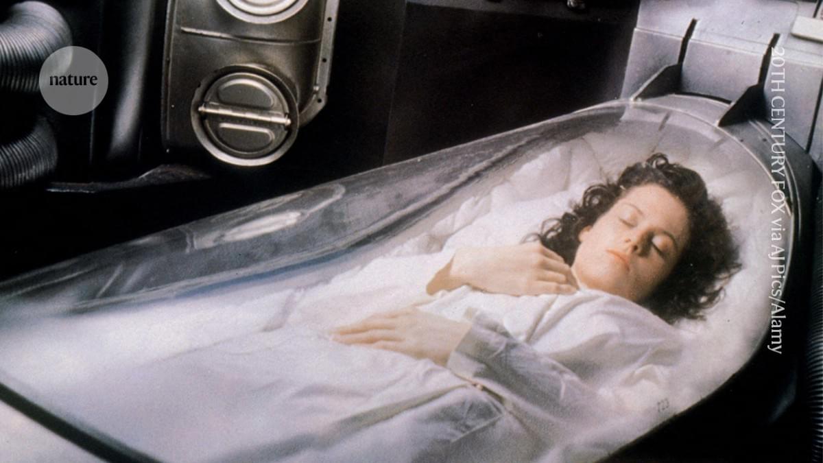
A familiar trope in science fiction is the cryopreserved time traveller, their body deep-frozen in suspended animation, then thawed and reawakened in another decade or century with all of their mental and physical capabilities intact.

Researchers attempting the cryogenic freezing and thawing of brain tissue from humans and other animals — mostly young vertebrates — have already shown that neuronal tissue can survive freezing on a cellular level and, after thawing, a functional one to some extent. But it has not been possible to fully restore the processes necessary for proper brain functioning — neuronal firing, cell metabolism and brain plasticity.

A team in Germany has now demonstrated a method for cryopreserving and thawing mouse brains that leaves some of this functionality intact. The study, published on 3 March in Proceedings of the National Academy of Sciences 3, details the authors’ use of a method called vitrification, which preserves tissue in a glass-like state, along with a thawing process that preserves living tissue.

“If brain function is an emergent property of its physical structure, how can we recover it from complete shutdown?” asks Alexander German, a neurologist at the University of Erlangen–Nuremberg in Germany and lead author of the study. The findings, he says, hint at the potential to one day protect the brain during disease or in the wake of severe injury, set up organ banks and even achieve whole-body cryopreservation of mammals.

Mrityunjay Kothari, who studies mechanical engineering at the University of New Hampshire in Durham, agrees that the study advances the state of the art in cryopreservation of brain tissue. “This kind of progress is what gradually turns science fiction into scientific possibility,” he says. However, he adds that applications such as the long-term banking of large organs or mammals remain far beyond the capabilities of the study.

Article Featured in Nature.


Compact terahertz imaging system brings real-time, non-invasive clinical diagnostics closer

Scientists at the University of Warwick and University of Exeter have developed a fully fiber-coupled terahertz (THz) imaging system that significantly improves the speed, resolution, and clinical practicality of terahertz imaging. The study, published in Nature Communications, demonstrates a high-throughput, compact platform that overcomes key barriers limiting current THz systems—bringing real-time, non-invasive tissue imaging closer to routine clinical use.

“Terahertz imaging has shown immense promise for biomedical diagnostics, but its translation into real-world clinical tools has been hindered by bulky systems and slow acquisition speeds,” said Professor Emma MacPherson, Department of Physics, University of Warwick. “It’s an exciting breakthrough as the fiber coupling means that the system can be flexible and compact, meaning it can function as a handheld device or be integrated with a robot.”

Terahertz waves sit between microwaves and infrared light on the electromagnetic spectrum. Crucially, they are non-ionizing (meaning they do not carry the risks associated with X-rays) and are highly sensitive to water content, which helps reveal differences between healthy and diseased tissue. Despite this promise, most existing terahertz imaging systems are bulky and slow, limiting their use outside specialist labs.

/* */