Toggle light / dark theme

How whales defy the cancer odds: Good genes

But they don’t. Instead, they are less likely to develop or die of this enigmatic disease. The same is true of elephants and dinosaurs’ living relatives, birds. Marc Tollis, an assistant professor in the School of Informatics, Computing, and Cyber Systems at Northern Arizona University, wants to know why.

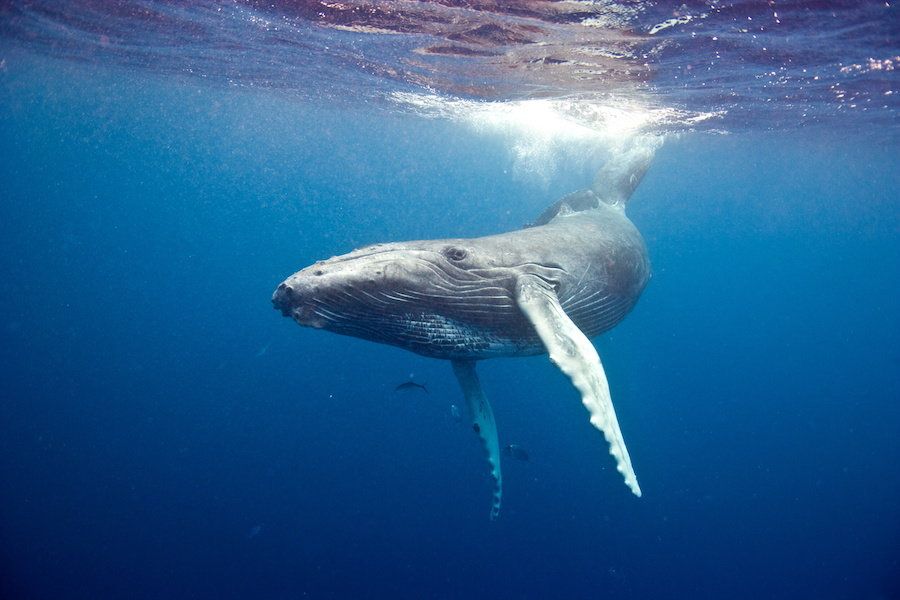
Tollis led a team of scientists from Arizona State University, the University of Groningen in the Netherlands, the Center for Coastal Studies in Massachusetts and nine other institutions worldwide to study potential cancer suppression mechanisms in cetaceans, the mammalian group that includes whales, dolphins and porpoises. Their findings, which picked apart the genome of the humpback whale, as well as the genomes of nine other cetaceans, in order to determine how their cancer defenses are so effective, were published today in Molecular Biology and Evolution.

The study is the first major contribution from the newly formed Arizona Cancer and Evolution Center or ACE, directed by Carlo Maley under an $8.5 million award from the National Cancer Institute. Maley, an evolutionary biologist, is a researcher at ASU’s Biodesign Virginia G. Piper Center for Personalized Diagnostics and professor in the School of Life Sciences. He is a senior co-author of the new study.


Europe abandons plans for ‘flagship’ billion-euro research projects

The consortium, called LifeTime, aims to use three emerging technologies—machine learning, the study of single cells, and lab-grown organlike tissues called organoids—to map how human cells change over time and develop diseases. It is one of six candidates in the latest round of ambitious proposals for European flagships, billion-euro research projects intended to run for 10 years. There is just one snag: The European Commission has decided that it won’t launch any of them.


Six candidate research proposals lost in limbo.

Read more

Boosting Levels of NAD+ May Make Senescent Cells More Aggressively Inflammatory

On senescent cells, mechanism of interaction and (potential?!) side effects of NAD+ supplements. “… Those people self-experimenting with NAD+ precursor supplementation should consider keeping a close eye on markers of inflammation…”


Enhancing levels of NAD+ in mitochondria via delivery of various precursor compounds as supplements is growing in popularity as an approach to boost faltering mitochondrial function and thus modestly slow the progression of aging. A human trial demonstrated improved vascular function as a result of nicotinamide riboside supplementation, for example. Researchers here show that increased NAD+ will likely make worse the inflammatory signaling of senescent cells, however. Senescent cells accumulate with age, and are an important cause of the chronic inflammation of aging that drives the progression of many age-related diseases.

The results here suggest that efficient senolytic treatments to selectively destroy senescent cells should proceed any of the current approaches to raising levels of NAD+ in older individuals — and it is an open question as to whether any of the existing available options are efficient enough to make NAD+ enhancement safe in the longer term. Those people self-experimenting with NAD+ precursor supplementation should consider keeping a close eye on markers of inflammation.

Read more

For The First Time, Scientists Turn Human Stem Cells Into Insulin-Producing Cells

May have posted this, but this is very cool. “We can now generate insulin-producing cells that look and act a lot like the pancreatic beta cells you and I have in our bodies,” explains one of the team, microphysiologist Matthias Hebrok from the University of California San Francisco (UCSF).


Although treatment of type 1 diabetes has come a long way since it was first described in Ancient Egypt, insulin injections and finger pricks are a daily part of life for many diabetics.

But researchers have just made a breakthrough that might one day make these technologies obsolete, by transforming human stem cells into functional insulin-producing cells (also known as beta cells) – at least in mice.

“We can now generate insulin-producing cells that look and act a lot like the pancreatic beta cells you and I have in our bodies,” explains one of the team, microphysiologist Matthias Hebrok from the University of California San Francisco (UCSF).

Read more

How stressed-out gut bacteria may trigger autoimmune response

Stressful life events most likely contribute to autoimmune diseases, but scientists don’t have a deep understanding of the underlying chain of events. A study on mice published this week in mSystems suggests that the gut microbiota may play a significant role in that connection. Researchers found that the onset of stress caused changes in the intestinal bacteria that, in turn, stimulated the activity of immune cells in a way that increased the likelihood that the body would a…


Chronic social stress in mice induces the expression of virulent genes in the gut microbiota. The altered microbiota increases the presence of effector T helper cells in the lymph nodes and induces myelin autoreactive cells. Exposure to chronic stress, therefore, may increase the risk of developing autoimmune diseases for some individuals with a susceptibility.

Read more

/* */