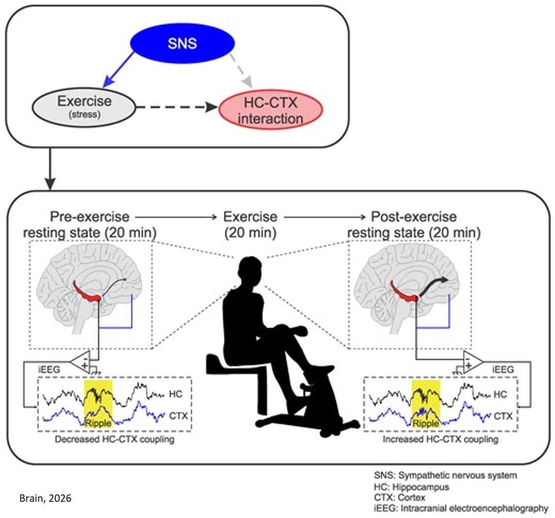
Toggle light / dark theme

Damage to the mitochondria, the “power plants” of the cells, contributes to many diseases. Researchers from Heinrich Heine University Düsseldorf (HHU) and the University of Cologne led by HHU professor of medicine Dr David Pla-Martín, now describe in the scientific journal Science Advances how cells with defective mitochondria activate a special recycling system to eliminate damaged genetic material.

Damage to the genetic material of mitochondria – the mitochondrial DNA or mtDNA for short – can lead to diseases such as Parkinson’s, Alzheimer’s, amyotrophic lateral sclerosis (ALS), cardiovascular diseases and type 2 diabetes. Such damage also speeds up the ageing process. However, the cells are normally capable of identifying such damage and reacting.

Damage to the genetic material of mitochondria—the mitochondrial DNA or mtDNA for short—can lead to diseases such as Parkinson’s, Alzheimer’s, amyotrophic lateral sclerosis (ALS), cardiovascular diseases and type 2 diabetes. Such damage also speeds up the aging process. However, the cells are normally capable of identifying such damage and reacting.

Scientists from University Hospital Düsseldorf and HHU have—in collaboration with the University of Cologne and the Center for Molecular Medicine Cologne (CMMC)—discovered a mechanism which protects and repairs the mitochondria. The research team, headed by Professor Pla-Martín from the Institute of Biochemistry and Molecular Biology I at HHU, has identified a specialized recycling system, which cells activate when they identify damage to the mtDNA.

According to the authors in Science Advances, this mechanism relies on a known as retromer and the lysosomes—cell organelles containing digestive enzymes. These special cellular compartments act like recycling centers, eliminating the damaged genetic material.

Most people have right-dominant hearts—which to a doctor or a researcher means they have an artery that extends from the right side of their hearts to supply oxygenated blood to the back side. For some people, this artery, called the posterior descending artery, comes from the left side or from both directions. A study has found that the gene CXCL12 is connected to this artery’s formation and that its directional pattern is set very early in human development.

The findings, reported in the journal Cell, represent a step toward developing “medical revascularization,” a long-term goal of Stanford researchers to create a treatment for blocked or limited-flow arteries by growing new ones to compensate.

“For the first time, we have evidence of a gene that regulates the development of one of the most important types of arteries in the human body,” said Kristy Red-Horse, co-senior author of the study and biology professor in the Stanford School of Humanities and Sciences. “And if we know the development pathways of these important arteries, then we can perhaps regrow them by reintroducing these pathways in a diseased heart.”

Training the brain’s immune system to recognize and clear toxic material is rapidly emerging as an promising way to put the brakes on Alzheimer’s disease. Unfortunately researchers haven’t been clear on how this method of protection operates on a cellular level.

An international team of researchers analyzed brain samples taken from people who had died with Alzheimer’s, some of whom had also received approved Alzheimer’s immunotherapy treatments. The therapies encourage cleaning cells called microglia to attack the clumps of amyloid-beta proteins that are thought to be involved in neurodegeneration.

Microglia responses to amyloid beta can lead to inflammation, which in turn risks damage to brain tissues. The researchers wanted to know why immunotherapy turned microglia into ruthless cleaning machines in some cases but not others.

Xueli Bai & team report on the high level of lactate in pancreatic cancer metabolism, which changes tumor immune features by protein lactylation and results in immunotherapy resistance:

The figure shows lactylation labeling of metabolic proteins from paracancerous and tumor tissues.


1Department of Hepatobiliary and Pancreatic Surgery, The First Affiliated Hospital, School of Medicine and.

2Zhejiang Provincial Key Laboratory of Pancreatic Disease, The First Affiliated Hospital, School of Medicine, Zhejiang University, Hangzhou, Zhejiang, China.

3MOE Joint International Research Laboratory of Pancreatic Diseases, Hangzhou, China.

R01 CA203108, R01 CA247234 (to ML), and by the William and Ella Owens Medical Research Foundation (to ML). It was also supported in part by the Department of Medicine, the University of Oklahoma Health Sciences Center.

Address correspondence to: Michael S. Bronze, Department of Medicine, The University of Oklahoma Health Sciences Center, 800 Stanton L. Young Blvd. AAT 6,400, Oklahoma City, Oklahoma, 73,104, USA. Phone: 405.271.5428; Email: [email protected]. Or to: Min Li, Department of Medicine, The University of Oklahoma Health Sciences Center, 975 NE 10th Street, BRC 1262A, Oklahoma City, Oklahoma, 73,104, USA. Phone: 405.271.1796; Email: [email protected].

Hedonic eating is defined as food consumption driven by palatability without physiological need. However, neural control of palatable food intake is poorly understood. We discovered that hedonic eating is controlled by a neural pathway from the peri–locus ceruleus to the ventral tegmental area (VTA). Using photometry-calibrated optogenetics, we found that VTA dopamine (VTADA) neurons encode palatability to bidirectionally regulate hedonic food consumption. VTADA neuron responsiveness was suppressed during food consumption by semaglutide, a glucagon-like peptide receptor 1 (GLP-1R) agonist used as an antiobesity drug. Mice recovered palatable food appetite and VTADA neuron activity during repeated semaglutide treatment, which was reversed by consumption-triggered VTADA neuron inhibition.