Toggle light / dark theme

Abstract: Unraveling TIME: CD8+ T cell-and CXCL11-driven endocrine resistance in BreastCancer:

Unraveling TIME: CD8+ T cell-and CXCL11-driven endocrine resistance in BreastCancer:

Tim Kong & Cynthia X. Ma provide a Commentary on Fabiana Napolitano: https://doi.org/10.1172/JCI188458


1Division of Oncology, Department of Medicine, Washington University School of Medicine, St. Louis, Missouri, USA.

2Department of Medicine, Weill Cornell Medicine, New York, New York, USA.

Address correspondence to: Cynthia X. Ma, Division of Oncology, Department of Medicine, Washington University School of Medicine, 660 South Euclid Avenue, St. Louis, Missouri, 63,110, USA. Email: [email protected].

AI tool can predict which trauma patients need blood transfusions before they reach the hospital

Severe bleeding is one of the most common and preventable causes of death after traumatic injury, yet currently available tools have poor ability to determine which patients urgently need blood transfusions. A new multinational study, just published in Lancet Digital Health, suggests artificial intelligence (AI) may help close that gap.

Researchers have developed and validated machine-learning models that can accurately predict whether trauma patients will require blood transfusions, using only information available before they reach the hospital such as vital signs, injury patterns, and medication history.

Co-author Prof Patricia Maguire from University College Dublin (UCD), Director of UCD AI Healthcare Hub and UCD Institute for Discovery, said, “These findings show that AI-driven decision support could enable earlier and more precise identification of patients at highest risk of hemorrhagic shock, using data already available to emergency services. This has clear potential to support more timely transfusion decisions, although prospective evaluation will be needed before clinical implementation.”

A Hidden Cellular Defense May Protect the Brain From Alzheimer’s

Scientists discovered why some neurons resist tau toxicity, identifying CRL5SOCS4 as a crucial defense and linking mitochondrial stress to harmful tau fragments. New research by UCLA Health and UC San Francisco has uncovered why certain brain cells are more resilient than others to the buildup of

Live-cell tracking reveals dynamic interaction between protein folding helpers and newly produced proteins

Proteins are the molecular machines of cells. They are produced in protein factories called ribosomes based on their blueprint—the genetic information. Here, the basic building blocks of proteins, amino acids, are assembled into long protein chains. Like the building blocks of a machine, individual proteins must have a specific three-dimensional structure to properly fulfill their functions.

To achieve this, the newly produced protein chains in human cells are folded into their stable and functional form with the help of various protein folding helper proteins, known as chaperones, such as TRiC/PFD, or HSP70/40. The protein folding helpers isolate the amino acid chains, which have different chemical properties depending on the amino acid, from the cellular environment. This prevents the newly produced protein chains from clumping together and causing disease.

F.-Ulrich Hartl, a director at the Max Planck Institute of Biochemistry, has spent decades studying the mechanisms of protein folding. Niko Dalheimer, a scientist in Hartl’s department and one of the two lead authors of a new study published in Nature, explains: Much of what we know about protein folding has been learned from studies conducted in test tubes. However, it is virtually impossible to faithfully replicate the cellular environment in vitro.

Faster enzyme screening could cut biocatalysis bottlenecks in drug development

A team of biochemists at the University of California, Santa Cruz, has developed a faster way to identify molecules in the lab that could lead to more effective pharmaceuticals. The discovery advances the rapidly growing field of biocatalysis, which depends on generating large, genetically diverse libraries of enzymes, and then screening those variants to find ones that perform a desired chemical task best.

This strategy has attracted major investment, particularly from drugmakers, because it promises quicker routes to complex, high-value molecules. However, traditional approaches to finding new biologically beneficial molecules often require “lots of shots on goal,” where researchers test enormous numbers of candidates through slow and inefficient workflows.

The method developed by the UC Santa Cruz team aims to significantly shorten that process by introducing smarter and faster decision-making tools that help researchers identify promising enzyme variants much earlier. The researchers detail their new approach in the journal Cell Reports Physical Science.

CRISPR screen maps 250 genes essential for human muscle fiber formation

Muscles make up nearly 40% of the human body and power every move we make, from a child’s first steps to recovery after injury. For some, however, muscle development goes awry, leading to weakness, delayed motor milestones or lifelong disabilities. New research from the University of Georgia is shedding light on why.

UGA researchers have created a first-of-its-kind CRISPR screening platform for human muscle cells, identifying hundreds of genes critical to skeletal muscle formation and uncovering the potential cause of a rare genetic disorder. The findings come from two companion papers published in Nature Communications, one describing the large-scale screen and a second digging into a particular gene’s role in muscle development.

Together, the studies provide a comprehensive genetic map of how human muscle fibers are built and lend insights into the effects of genetic mutations on developmental muscle defects. By linking specific genes to the muscle-building process, this genetic roadmap gives clinicians a practical shortlist to more quickly pinpoint the likely genetic causes of a patient’s muscle-development disorder. It also provides researchers with clear targets to prioritize future drug or gene therapy approaches.

Surgery for quantum bits: Bit-flip errors corrected during superconducting qubit operations

Quantum computers hold great promise for exciting applications in the future, but for now they keep presenting physicists and engineers with a series of challenges and conundrums. One of them relates to decoherence and the errors that result from it: bit flips and phase flips. Such errors mean that the logical unit of a quantum computer, the qubit, can suddenly and unpredictably change its state from “0” to “1,” or that the relative phase of a superposition state can jump from positive to negative.

These errors can be held at bay by building a logical qubit out of many physical qubits and constantly applying error correction protocols. This approach takes care of storing the quantum information relatively safely over time. However, at some point it becomes necessary to exit storage mode and do something useful with the qubit—like applying a quantum gate, which is the building block of quantum algorithms.

The research group led by D-PHYS Professor Andreas Wallraff, in collaboration with the Paul Scherrer Institute (PSI) and the theory team of Professor Markus Müller at RWTH Aachen University and Forschungszentrum Jülich, has now demonstrated a technique that makes it possible to perform a quantum operation between superconducting logical qubits while correcting for potential errors occurring during the operation. The researchers have just published their results in Nature Physics.

Next-generation immune profiling — beyond blood cancer cells

Why immunoscores work in solid tumors—but not yet in blood cancers👇

✅In solid tumors, immune profiling has reached a high level of standardization. Clear tumor boundaries allow quantification of immune cell infiltration, particularly CD3⁺ and CD8⁺ T cells, using immunohistochemistry. This has led to the development of validated immunoscores that stratify tumors as “hot,” “cold,” or “very cold,” providing robust prognostic and predictive value for immunotherapy response.

✅These immunoscores work because solid tumors are spatially organized. Immune cells can be classified as infiltrating or excluded, and their density within defined tumor regions directly correlates with clinical outcome. As a result, immune cell infiltration has become a reliable biomarker to guide treatment decisions in cancers such as colon carcinoma.

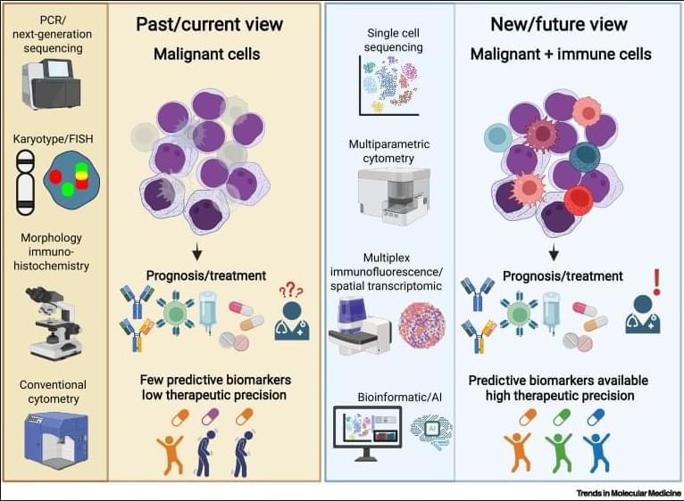
✅In contrast, hematologic malignancies lack these defining features. Leukemias and lymphomas are systemic diseases without clear tumor borders, making spatial immune assessment fundamentally challenging. Malignant and nonmalignant immune cells coexist within the same compartments, blurring the distinction between tumor cells and the immune microenvironment.

✅Current immune profiling in hematologic cancers relies on baseline physiological levels of circulating or tissue-resident immune cells, including monocytes, neutrophils, T cells, NK cells, and B cells. While techniques such as flow cytometry, histology, and bulk or single-cell RNA sequencing provide rich datasets, they do not yet translate into a unified, clinically actionable immune score.

✅This lack of standardization creates uncertainty in predicting immunotherapy responses. Metrics such as inflammation, cytotoxicity, or immune infiltration are difficult to interpret consistently across patients and disease subtypes, especially given systemic involvement and tissue-specific immune contexts.

💡

/* */