Toggle light / dark theme

Immune gene diversity and STING1 variants in shaping cancer immunity across different genetic ancestry populations

Hu et al. analyzed non-synonymous SNPs across diverse human populations and revealed divergent evolutionary pressures on immune-and cancer-related genes. By integrating population diversity with functional evaluation, they identified STING1 variants as modulators of interferon signaling. Their findings suggest that germline variations shaped by genetic ancestry may influence cancer immunity.

PRISM reanalyzes 4,400 tumors, reshaping the debate over tumor microbiomes

When scientists sequence tumor DNA, they typically find small amounts of genetic code from bacteria, viruses and fungi—microorganisms that—if actually present in tumor tissues—could influence how they grow, evade immunity or respond to treatment. But do microorganisms truly reside in tumors, or do the samples become contaminated before sequencing occurs?

Independent analyses of the same genomic data have reached wildly different conclusions. Now, researchers at Rutgers Cancer Institute have developed a computational tool that settles the controversy by distinguishing genuine microbial signals from artifacts. Their findings are published in Cancer Cell.

“There are microbes all over the environment, on our skin and in our breath,” said Subhajyoti De, a member of the Genomic Instability and Cancer Genetics Program at Rutgers Cancer Institute and the senior author of the study. “There could be DNA particles floating in the air. How do you know whether you’re finding came from the tissue you were interested in, or whether something was introduced along the way?”

Ultrastructural and Histological Cryopreservation of Mammalian Brains by Vitrification

Studies of whole brain cryopreservation are rare but are potentially important for a variety of applications. It has been demonstrated that ultrastructure in whole rabbit and pig brains can be cryopreserved by vitrification (ice-free cryopreservation) after prior aldehyde fixation, but fixation limits the range of studies that can be done by neurobiologists, including studies that depend upon general molecular integrity, signal transduction, macromolecular synthesis, and other physiological processes. We now show that whole brain ultrastructure can be preserved by vitrification without prior aldehyde fixation. Rabbit brain perfusion with the M22 vitrification solution followed by vitrification, warming, and fixation showed an absence of visible ice damage and overall structural preservation, but osmotic brain shrinkage sufficient to distort and obscure neuroanatomical detail. Neuroanatomical preservation in the presence of M22 was also investigated in human cerebral cortical biopsies taken after whole brain perfusion with M22. These biopsies did not form ice upon cooling or warming, and high power electron microscopy showed dehydrated and electron-dense but predominantly intact cells, neuropil, and synapses with no signs of ice crystal damage, and partial dilution of these samples restored normal cortical pyramidal cell shapes. To further evaluate ultrastructural preservation within the severely dehydrated brain, rabbit brains were perfused with M22 and then partially washed free of M22 before fixation. Perfusion dilution of the brain to 3-5M M22 resulted in brain re-expansion and the re-appearance of well-defined neuroanatomical features, but rehydration of the brain to 1M M22 resulted in ultrastructural damage suggestive of preventable osmotic injury caused by incomplete removal of M22. We conclude that both animal and human brains can be cryopreserved by vitrification with predominant retention of ultrastructural integrity without the need for prior aldehyde fixation. This observation has direct relevance to the feasibility of human cryopreservation, for which direct evidence has been lacking until this report. It also provides a starting point for perfecting brain cryopreservation, which may be necessary for lengthy space travel and could allow future medical time travel.

The authors have declared no competing interest.

What to watch as fungal infections rise: Species that can quickly ‘translate’ fat-use proteins

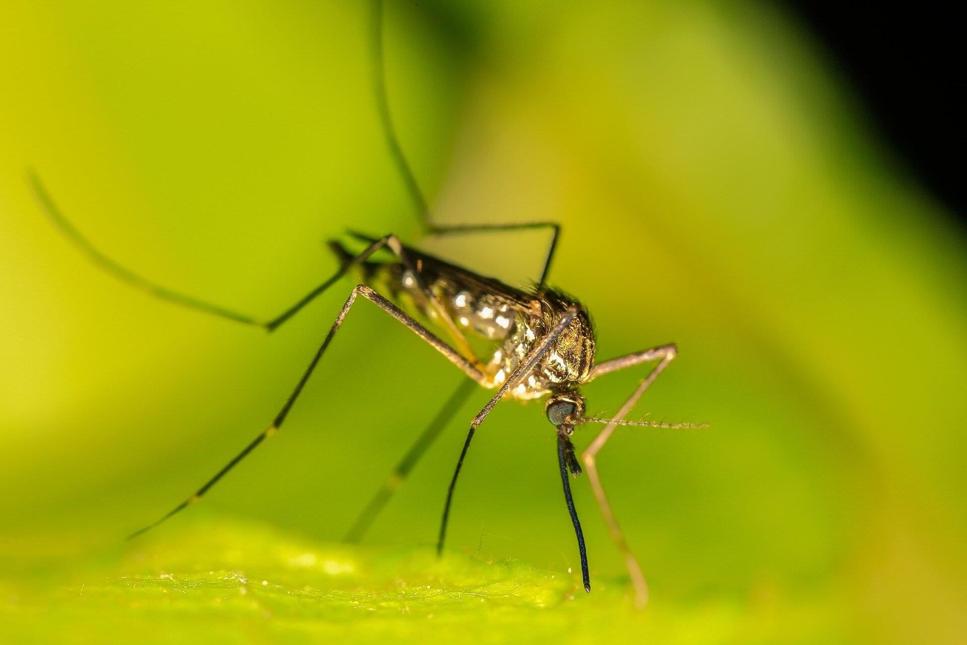
A new study by researchers at Kiel University and MPI-EvolBio describes how more efficient protein production drives the adaptation of fungi to the human body, potentially turning previously harmless species into emerging pathogens. In the wake of global change and the associated rise in temperatures, fungal infections are on the increase worldwide, threatening crops, wildlife and, also, human health. Many fungal species are completely harmless and fulfill important ecological functions, such as decomposing organic matter and releasing nutrients into the soil.

As symbionts of multicellular organisms, they perform useful functions for their host. On the other hand, some species are so-called opportunistic human pathogens: particularly in a weakened immune system, such fungi can colonize the body and cause serious and even life-threatening infections.

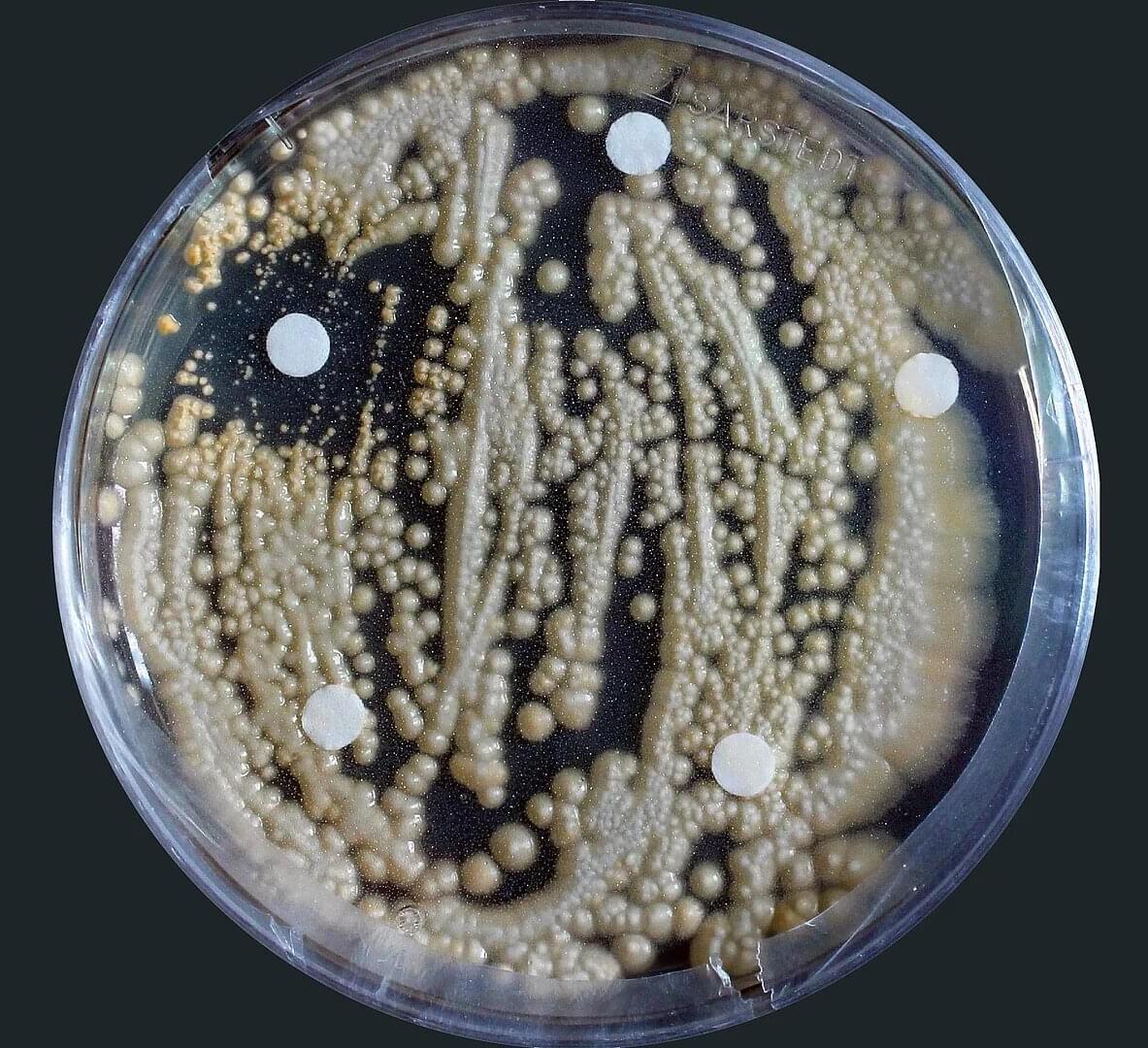
While fungi are often studied as pathogens of crops at institutions such as Kiel University and the Max Planck Institute for Evolutionary Biology in Plön (MPI-EvolBio), researchers are increasingly turning their attention to their harmful effects on humans. A research team led by Professor Eva Stukenbrock, head of the Environmental Genomics group at Kiel University and MPI-EvolBio, has conducted a new study to investigate why certain fungi might become human pathogens in the course of global change. To this end, the researchers analyzed various fungal species of the order Trichosporonales, which includes both harmless and dangerous species for humans.

GeSn alloys emerge as a new semiconductor class that could reshape optoelectronics

Scientists have created a new type of material that could enable common electronic devices to work faster and use less energy, a study suggests. The findings indicate the material, which was until now thought near-impossible to make, can act as a highly effective semiconductor—a key component of modern electrical devices.

Using the new semiconductor in electronics such as computer processors or medical imaging devices could help them run more efficiently, the team says.

Indigenous Food Is Medicine Program Feasibility for Navajo Patients

In this nonrandomized clinical trial, the MUTTON-HF intervention incorporating Indigenous recipes and locally sourced Native food was feasible and acceptable for patients with heart failure in rural Navajo Nation.


Question Among Navajo patients with heart failure living rurally on the reservation, is a medically tailored meal delivery program incorporating Indigenous foods and recipes feasible and acceptable?

Findings This nonrandomized clinical trial included 20 American Indian patients with heart failure receiving care at 2 Indian Health Service sites in rural Navajo Nation. A community-designed, Indigenous, medically tailored meal program was implemented; the intervention was deemed both feasible (90% of weekly meal boxes received by patients) and acceptable (mean Acceptability of Intervention Measure score, 17 of 20).

Development of human induced pluripotent stem cell-derived ovarian support cells as a clinical-grade product for in vitro fertilization

Paulsen et al. present the process development and clinical application of an hiPSC-derived OSC product, Fertilo. They describe the raw material upgrades, process consistency and reproducibility, and analytical assessment required for the generation of a clinically suitable product, as well as favorable outcomes from the first-in-human application of Fertilo.

Scientists now know why ovarian cancer spreads so rapidly in the abdomen

Ovarian cancer kills more women than any other gynecological cancer. Most patients receive their diagnosis only after the disease spreads throughout the abdomen. Until now, scientists have never fully understood why this cancer advances so fast.

A new study led by Nagoya University explains why. Published in Science Advances, the study shows that cancer cells recruit help from protective mesothelial cells that normally line the abdominal cavity. Mesothelial cells lead the invasion and cancer cells follow the pathways they create. These hybrid cell clusters resist chemotherapy better than cancer alone.

Researchers examined abdominal fluid from ovarian cancer patients and found something unexpected. Cancer cells do not float alone in the abdominal cavity. Instead, they often grab onto mesothelial cells and form hybrid spheres. About 60% of all cancer spheres contain these recruited mesothelial cells. The cancer cells release a protein called TGF-β1 that transforms the mesothelial cells and causes them to develop spike-like structures that cut through tissue.

ABCA1 protein releases molecular brakes on solid tumor immunotherapy, study finds

In recent years, cancer researchers have made major breakthroughs by using the body’s immune system to fight cancer. One of the most promising approaches, known as immune checkpoint blockade, works by releasing molecular “brakes” on T cells. This allows them to better recognize and attack cancer cells. While these therapies can be very effective for some patients, many solid tumors, including most forms of breast cancer, remain largely unaffected. Cancer Center at Illinois (CCIL) Program Co-leader Erik Nelson and his research group are working to understand why these treatments fail.

Elevated blood concentrations of cholesterol have long been linked to cancer outcomes. In a new study, they found that a protein called ABCA1 is involved in transporting cholesterol out of a type of immune cell called macrophages, and in so-doing shifts them to an “attack cancer” mode.

“Immune based therapies have revolutionized how we can treat cancer, basically taking the brakes off of a type of immune cell called T cells so they can attack cancer,” Nelson said. “While this approach works well for some patients, many so-called solid tumors fail to respond or develop resistance mechanisms.”

DNA marker in malaria mosquitoes may be pivotal in tackling insecticide resistance

A new study has detected a DNA marker in a gene encoding a key enzyme known as cytochrome P450 that helps mosquitoes to break down and survive exposure to pyrethroids, the main insecticides used for treating bed nets. This new finding, published on the bioRxiv preprint server and slated for publication in Science Translational Medicine, will help to better implement insecticide resistance management strategies and contribute to reducing the burden of malaria in sub-Saharan Africa, home to 90% of cases globally.

The work was jointly led by Liverpool School of Tropical Medicine and the Centre for Research in Infectious Diseases (CRID) in Cameroon.

Professor Charles Wondji, Professor of Genetics and Vector Biology at Liverpool School of Tropical Medicine and lead author on the study, said, “Our study designed field-applicable tools to easily track the spread of metabolic resistance in the major malaria mosquito species and assess its impact on control interventions. These important findings can help to maintain the effectiveness of insecticide-based tools such as bed nets, which remain a cornerstone of malaria prevention.”

/* */