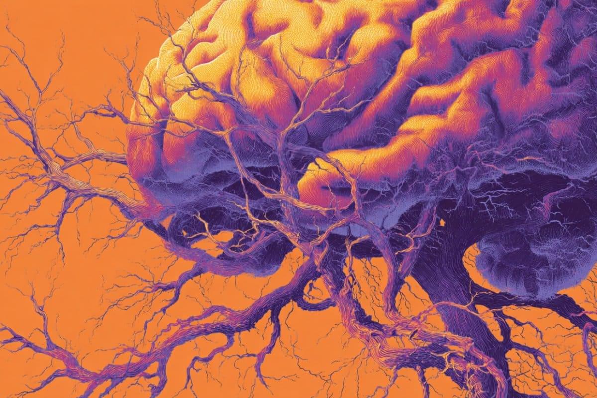
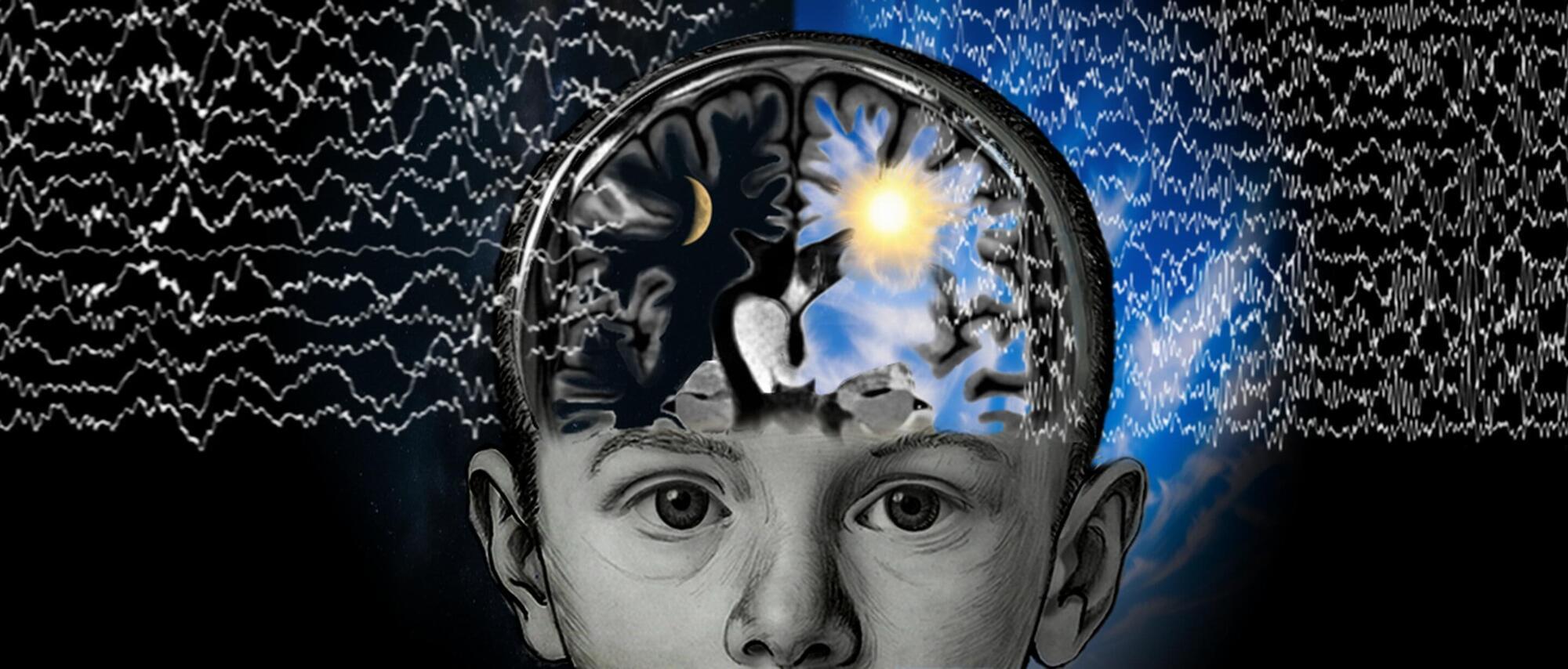
A new study reveals that sleep-like slow-wave brain activity can persist for years in surgically disconnected brain hemispheres of awake epilepsy patients.


EPFL researchers have demonstrated the first pill-sized bioprinter that can be swallowed and guided within the gastrointestinal tract, where it directly deposits bio-ink over damaged tissues to support repair.
Soft tissue injuries of the gastrointestinal tract, like ulcers or hemorrhages, can currently be treated only with some form of surgery, which is invasive and may not result in permanent repair. Bioprinting is emerging as an effective treatment that deposits biocompatible “ink”—often made of natural polymers derived from seaweed—directly over the site of tissue damage, creating a scaffold for new cell growth. But like traditional surgical tools, these kinds of bioprinters tend to be bulky and require anesthesia.
At the same time, “untethered” technologies are being developed to perform medical interventions without a physical connection to external equipment. For example, ingestible “smart capsules” can be guided to drug delivery sites using external magnets. But these devices are designed to travel through liquids, and their movements become unpredictable when they touch the tissue wall.

face_with_colon_three Year 1998
In ancient Greece, immortality was the province of gods who spun the length of each lifetime. The myth has a kernel of truth, because the ends of chromosomes are protected by specialized stretches of DNA called telomeres. Once these are snipped too much by imperfect copying, a cell goes into senescence and stops dividing. Now two reports show that, with the help of an enzyme called telomerase, human cells can divide forever in the laboratory without turning cancerous. The findings, reported in the January issue of Nature Genetics, could ease the way to new treatments for burn victims, diabetics, and patients with other diseases.
Researchers hoped that adding telomerase would keep cells dividing long enough to replace tissues lost to injury or disease. Normal cells often have proved impractical because they can only divide a limited number of times in culture, and once returned to the body they’re often too old to do much good. The limitation may be that normal cells do not produce active telomerase, which can rebuild the telomeres and keep cells from becoming senescent.
In fact, about a year ago, Jerry Shay and his colleagues at the University of Texas Southwestern Medical Center in Dallas showed that adding the enzyme to normal connective tissue cells called fibroblasts extends their life-span (Science NOW, 13 January 1998). These cells have now lived three times longer than normal in the lab, and they are still going strong. But because cancer cells contain telomerase and also live forever, scientists worried that the newly immortal cells would become malignant when implanted in humans.

Cancer is classified as the uncontrollable growth of mutated cells. There are different types of cancers that correlate to various tissues and organs. When individuals hear “cancer” they tend to think of breast cancer or another type of solid tumor. While about 90% of new cancer diagnoses are solid tumors, the remaining 10% are hematologic or blood cancers. Hematological malignancies affect the blood, bone marrow, and lymph nodes. Specific blood cancers include leukemia, lymphoma (Hodgkin’s and non-Hodgkin’s), and multiple myeloma. Around the world, these cancers account for about 7% of all cancer-related deaths, with a projection to increase to about 4.6 million cases in 2030.
Symptoms for hematological malignancies can vary and present in a wide range. Most common symptoms include fatigue, fever, weight loss, bruising, bleeding easily, anemia, low platelet count, and low white blood count. Many patients will also feel bone pain and muscle weakness accompanied by headaches and seizures. Current standard-of-care therapy include chemotherapy combined with another form of treatment. Dependent on the patient and the stage of the cancer, physicians can prescribe targeted therapies, immunotherapies, and stem cell transplants. However, some cancers find ways to resist therapy and continue to progress. Currently, many scientists are working to overcome barriers and improve therapy for patients with hematological malignancies.
A recent article in Science Advances, by Dr. Philippe Bousso and others, demonstrated that an immunotherapy can elicit a strong antitumor response by reprogramming malignant immune cells in lymphomas and leukemias. Bousso is an immunologist and head of the Dynamics of Immune Responses Unit at the Pasteur Institute in France. His work focuses on understanding immune responses in different diseases using innovative imaging approaches. Importantly, his work has helped the field of immunology redefine immune cell functions and showed how proteins secreted by immune cells can have an effect in distal locations throughout the body.


In addition to strengthening the muscles, lungs, and heart, regular physical exercise also strengthens the immune system. This finding comes from a study of older adults with a history of endurance training, which involves prolonged physical activity such as long-distance running, cycling, swimming, rowing, and walking.
An international team of researchers analyzed the defense cells of these individuals and found that “natural killer” cells, which patrol the body against viruses and diseased cells, were more adaptable, less inflammatory, and metabolically more efficient.
The research, published in the journal Scientific Reports, investigated natural killer (NK) cells. NK cells are a type of white blood cell (lymphocyte) that can destroy infected and diseased cells, including cancer cells. They are at the forefront of the immune system because they detect and fight viruses and other pathogens. The researchers analyzed the cells of nine individuals with an average age of 64, divided into two groups: untrained and trained in endurance exercise.

In a major leap for cancer research, Google DeepMind and Yale University have unveiled an artificial intelligence system capable of uncovering new biological insights directly validated in living cells.
Announced on October 15, the new foundation model, C2S-Scale 27B, represents one of the largest and most sophisticated AI systems ever developed to study cellular behavior.
Built on Google’s Gemma family of models, it has generated a groundbreaking hypothesis about how cancer cells interact with the immune system—one that could reshape how future therapies are designed.

Sleep-like slow-wave patterns persist for years in surgically disconnected neural tissue of awake epilepsy patients, according to a study published in PLOS Biology by Marcello Massimini from Universita degli Studi di Milano, Italy, and colleagues.
The presence of slow waves in the isolated hemisphere impairs consciousness; however, whether they serve any functional or plastic role remains unclear.
Hemispherotomy is a surgical procedure used to treat severe cases of epilepsy in children. The goal of this procedure is to achieve maximal disconnection of the diseased neural tissue, potentially encompassing an entire hemisphere, from the rest of the brain to prevent the spread of seizures.

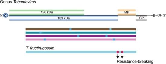
The genus Tobamovirus belongs to the family Virgaviridae, and the genome consists of monopartite, positive, single-strand RNA. Most species contain four open reading frames encoding four essential proteins. Transmission occurs primarily through mechanical contact between plants, and in some cases, via seed dispersal. Tobamovirus fructirugosum (tomato brown rugose fruit virus, ToBRFV), the most recently described species in the genus, was first reported in 2015. It overcame genetic resistance that had been effective in tomato for sixty years, causing devastating losses in tomato production worldwide, and highlights the importance of understanding Tobamovirus genomic variation and evolution. In this study, we measured and characterized nucleotide variation for the entire genome and for all species in the genus Tobamovirus.
