Toggle light / dark theme

Failing hearts nearly returned to full function in laboratory pigs after they received an experimental gene therapy.

New research shows the gene therapy didn’t just prevent heart failure from worsening in four lab pigs, but actually prompted hearts to repair and grow stronger.

“Even though the animals are still facing stress on the heart to induce heart failure, we saw recovery of heart function and that the heart also stabilizes or shrinks,” said co-senior researcher Dr. TingTing Hong, an associate professor of pharmacology and toxicology at the University of Utah.

Achieving the aggregation of different mutation types at multiple genomic loci and generating transgene-free plants in the T0 generation is an important goal in crop breeding. Although prime editing (PE), as the latest precise gene editing technology, can achieve any type of base substitution and small insertions or deletions, there are significant differences in efficiency between different editing sites, making it a major challenge to aggregate multiple mutation types within the same plant.

Recently, a collaborative research team led by Li Jiayang from the Institute of Genetics and Developmental Biology (IGDB) of the Chinese Academy of Science, developed a multiplex gene editing tool named the Cas9-PE system, capable of simultaneously achieving precise editing and site-specific random mutagenesis in rice.

By co-editing the ALSS627I gene to confer resistance to the herbicide bispyribac-sodium (BS) as a selection marker, and using Agrobacterium-mediated transient transformation, the researchers also achieved transgene-free gene editing in the T0 generation.

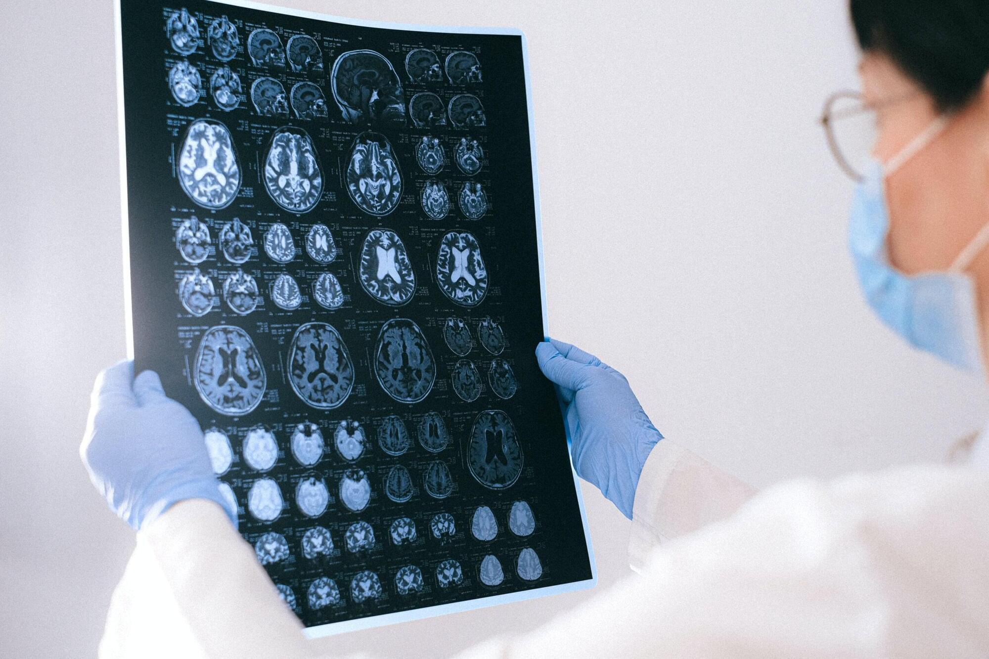
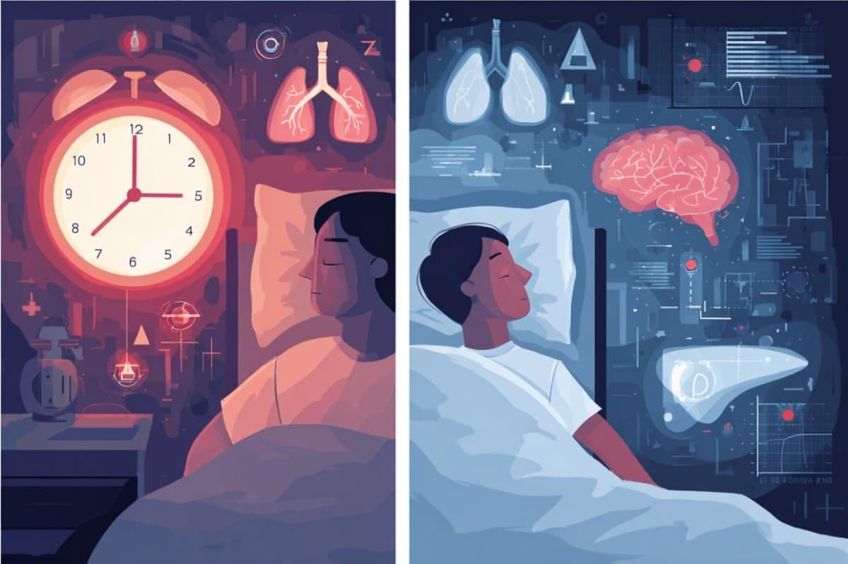
The shift from an awake state to unconsciousness is a phenomenon that has long captured the interest of scientists and philosophers alike, but how it happens has remained a mystery—until now. Through studies on rats, a team of researchers at Penn State has pinpointed the exact moment of loss of consciousness due to anesthesia, mapping what happens in different brain regions during that moment.

The study has implications for humans as well as for other types of loss of , such as sleep, the researchers said. They published their results in Advanced Science.

“People in the neuroscience field generally understand what happens to a patient who is going under anesthesia at a ,” said corresponding author Nanyin Zhang, the Dorothy Foehr Huck and J. Lloyd Huck Chair in Brain Imaging and professor of biomedical engineering at Penn State.

Summary: The dural sinuses and skull bone marrow serve as key communication hubs between the brain’s central immune system and the body’s peripheral immune system. These regions may act as “traffic lights,” allowing immune signals to flow between the brain and body, challenging the traditional view of the blood-brain barrier as an absolute divide.

Researchers found inflammatory activity in these areas correlates with inflammation in both the brain and body, offering new insights into conditions like depression. This discovery could pave the way for innovative treatments targeting these hubs to address immune-related conditions more precisely.

Therapeutic mRNAs offer great potential as a versatile and precise tool against cancer and other diseases. However, the therapeutic effectiveness is limited by the poor translation uptake of naked mRNA. To circumvent this challenge, researchers from VIB, VUB, Ghent University, and eTheRNA Immunotherapies developed an immunotherapeutic platform based on lipid-based nanoparticles (LNPs).

In different cancer models, applying a novel mixture of immunotherapeutic mRNA encapsulated in LNPs led to a clearly improved therapeutic efficacy with limited side effects. This proves the added value of the platform to the development of effective mRNA immunotherapies. The work is published in the journal Nature Communications.

The COVID-19 pandemic and recent Nobel Prize recognition have spotlighted mRNA therapies as a promising approach for diseases like cancer. With precision, scalability, and controlled , mRNA-based immunotherapy can encode proteins that stimulate the immune system to target and destroy cancer cells. Yet, naked mRNA is unstable, prone to degradation, and poorly absorbed by cells, limiting its effectiveness. This makes the development of reliable delivery methods essential for the future success of mRNA immunotherapies.

A new USC Stem Cell study published in the Proceedings of the National Academy of Sciences has identified key gene regulators that enable some deafened animals—including fish and lizards—to naturally regenerate their hearing. The findings could guide future efforts to stimulate the regeneration of sensory hearing cells in patients with hearing loss and balance disorders.

Led by first author Tuo Shi and co-corresponding authors Ksenia Gnedeva and Gage Crump at the Keck School of Medicine of USC, the study focuses on two cell types in the inner ear: the sensory cells that detect sound, and the that create an environment where sensory cells can thrive.

In highly regenerative species such as fish and lizards, supporting cells can also transform into replacement sensory cells after injury—a capacity absent in humans, mice and all other mammals.

A year later, he got a myoelectric arm, a type of prosthetic powered by the electrical signals in his residual limb’s muscles. But Smith hardly used it because it was “very, very slow” and had a limited range of movements. He could open and close the hand, but not do much else. He tried other robotic arms over the years, but they had similar problems.

“They’re just not super functional,” he says. “There’s a massive delay between executing a function and then having the prosthetic actually do it. In my day-to-day life, it just became faster to figure out other ways to do things.”

Recently, he’s been trying out a new system by Austin-based startup Phantom Neuro that has the potential to provide more lifelike control of prosthetic limbs. The company is building a thin, flexible muscle implant to allow amputees a wider, more natural range of movement just by thinking about the gestures they want to make.

How do human organs develop and what happens to them when they become diseased? To answer these questions, researchers are increasingly focusing on so-called organoids. These mini-organs, just a few millimeters in size, consist of groups of cells cultivated in the laboratory that can form organ-like structures.

Similar to embryonic development, organoids make it possible to investigate the interaction of cells in three-dimensional space—for example in metabolic processes or disease mechanisms.

The production of organoids is tricky; the required nutrients, and signaling molecules must be added in a specific order and at specific times according to a precise schedule.