Toggle light / dark theme

Yay face_with_colon_three


Researchers discovered that PLK1 triggers a process ensuring centromere preservation during cell division by activating the Mis18 complex and controlling CENP-A loading. This finding is key to understanding how cells correctly divide their genetic material, preventing diseases like cancer.

Scientists have resolved a decade-long mystery about the mechanism that maintains the centromere, the crucial region responsible for ensuring accurate DNA division during cell division.

A study revealed that a protein, known as PLK1, triggers a process that coordinates key proteins at the right place and time during cell division – ensuring each new cell has a centromere in the right location.

Intestinal stem cells play an important role in maintaining intestinal homeostasis and repairing damaged epithelial tissue. These cells function in a regenerative manner to generate new tissue throughout the growth phase and repair damaged tissue during the aging process.101 The interactions between the gut microbiome and intestinal stem cells are crucial because, if this interaction is comprehended, it may be possible to address various disorders that require stem cell therapy, heal wounds, and improve the durability of organ transplants.101 A recent study showed a connection between hematopoietic stem cells and the microbiome through altering metabolic stress.66 Therefore, the microbiota is crucial for maintaining microbial homeostasis, regulating metabolism, and the innate and adaptive immune systems.101 Furthermore, the study reveals that compositional alterations in the gut microbiome driven by dysbiosis are related to stem cell aging, metabolic dysregulations, stem cells’ epigenetic instability, and abnormal immune system activation.66

In the field of anti-aging, stem cells are regarded to have great potential. In numerous organs, it has been demonstrated that as we age, stem cells lose their capacity for self-renewal and differentiation and run out of resources.89 The emergence of anti-aging medications should address the dysregulation caused by aging that affects stem cells’ capacity for differentiation and self-renewal by re-regulating intrinsic and extrinsic variables. The host microbiome, hormones, local immune system, systemic inflammation, and niche structure are just a few examples of microenvironmental and systemic factors that influence stem cell aging.66

Endogenous ethanol is a class of microbiological metabolites. Proteobacteria, including E. coli and other Enterobacteriaceae, produce ethanol with bacterial origins. High endogenous ethanol levels in the human hippocampus inhibit proliferating stem cells and reduce progenitor and stem cells.102 Additionally, when more ethanol accumulates in the gut, it enhances the permeability of the gut by disrupting epithelial tight junctions, particularly zonula occludens. This enables the movement of pathogenic microbes, their endotoxins, and ethanol across the epithelial layer, causing more immediate and adverse effects on tissues. As a result, the stem cell reserve depletes, hastening the aging process and compensating for damaged tissues.103

Source: Nottingham Trent University.

Scientists have identified previously unreported genes which appear to play a key role in the muscle aging process. It is hoped that the findings from a Nottingham Trent University study could be used to help delay the impact of the aging process.

The study, which also involved Sweden’s Karolinska Institute, Karolinska University Hospital, and Anglia Ruskin University, is reported in the Journal of Cachexia, Sarcopenia and Muscle.

A new University of Maryland-led discovery could spur the development of new and improved treatments for Hutchinson-Gilford progeria syndrome (HGPS), often simply called “progeria”—a rare genetic disorder with no known cure that causes accelerated aging in children.

Publishing in the journal Aging…


Researchers identify protein that could improve cardiovascular health of those with progeria.

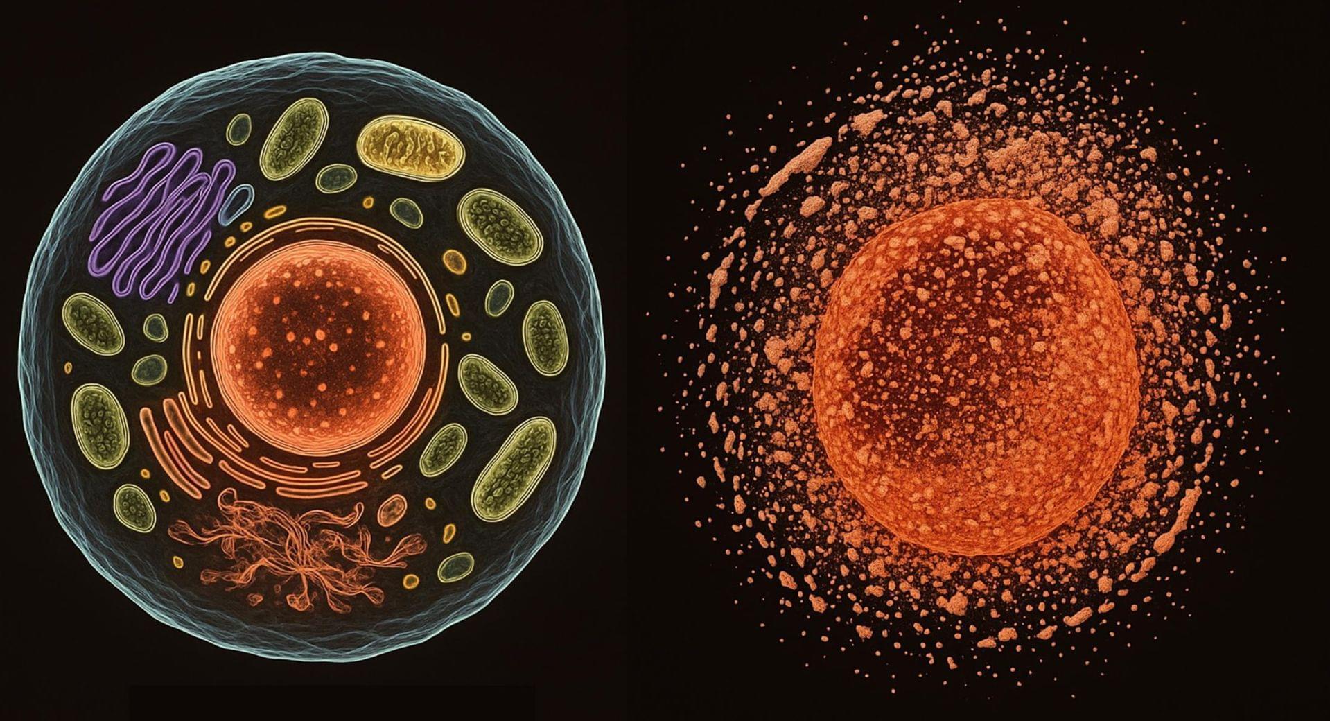
The research focuses on “cellular senescence,” a process where cells stop dividing and enter a state associated with chronic inflammation and aging.

This cellular state, known as the senescence-associated secretory phenotype (SASP), involves the secretion of inflammatory proteins that accelerate aging and disease, such as dementia, diabetes, and atherosclerosis.

Despite its almost perfect anti-aging profile, rapamycin exerts one significant limitation – inappropriate physicochemical properties. Therefore, we have decided to utilize virtual high-throughput screening and fragment-based design in search of novel mTOR inhibiting scaffolds with suitable physicochemical parameters. Seven lead compounds were selected from the list of obtained hits that were commercially available (4, 5, and 7) or their synthesis was feasible (1, 2, 3, and 6) and evaluated in vitro and subsequently in vivo. Of all these substances, only compound 3 demonstrated a significant cytotoxic, senolytic, and senomorphic effect on normal and cancerous cells. Further, it has been confirmed that compound 3 is a direct mTORC1 inhibitor. Last but not least, compound 3 was found to exhibit anti-SASP activity concurrently being relatively safe within the test of in vivo tolerability. All these outstanding results highlight compound 3 as a scaffold worthy of further investigation.

GRAPHICAL ABSTRACT