Toggle light / dark theme

Now, this is intriguing — pathways are a critical part of our system that monitors and manages how our bodies respond and interact to changes in our bodies. This recent SRC report focuses on the researchers efforts in monitoring pathways and how defects in pathways contribute to the biology and pathophysiology of cancer.


Bethesda, MD — This SRC focuses on new developments in the biology of lipid signaling with an emphasis on cancer, neuronal and cardiovascular diseases. The emphasis will be on molecular, cellular, structure/function and enzymatic mechanisms of physiological signaling pathways and how defects in these pathways contribute to the biology and pathophysiology of cancer, neurodegeneration and cardiovascular disease. The focus will be on how diacylglycerol, phosphatidic acid, lysophospholipids, sphingolipids and phosphoinositide lipids modulate specific pathways and processes in the contexts of physiological growth-regulatory signals, intracellular and extracellular vesicular trafficking, regulation of cell polarization, migration, motility and invasion, autophagy and epithelial extrusion, and as nuclear regulators of mRNA processing and gene expression. These sessions will include discussions on how signaling becomes dysfunctional in diseases. There will be presentations on new translational approaches and therapeutic targets. There will be significant representation from the pharmaceutical and biotechnology industry in order to facilitate networking between industry and academia. The topic areas have been chosen to maximize discussion of provocative and important developments.

We particularly wish to encourage the participation of new and junior researchers in the field and are securing additional support to provide PhD/postdoctoral fellow travel awards. Organizers have kept multiple short session speaking slots open. These will be selected from novel advances during 2015–2016 and from submitted abstracts. There will be multiple opportunities for new investigators and postdoctoral fellows to present and discuss their work including at poster sessions, short talks and short 5–10 minute oral ‘research snapshots’ to highlight their submitted abstracts. There will be multiple poster sessions during the conference. Time will also be allocated to at least two “meet the expert sessions” wherein established research leaders will dedicate time to interact with trainees and new investigators, specifically to give advice concerning the science and possible prospects for postdoctoral training, research funding, publishing or employment tracks.

The 2016 meeting brings together a wide range of leading investigators from across the globe. The scope of their subjects is vast, encompassing studies at the level of single proteins as well as the pathophysiology of complex disease. The program will highlight inter-disciplinary approaches and how major advances in biophysical, proteomic, genomic, imaging, modeling and therapeutic approaches are driving the field. The discussion forums and recreational activities will provide all participants extensive opportunities to exchange new ideas and forge new collaborations in a supportive interdisciplinary environment for participants at all stages of their research profession.

Finally. Bionic eye technology that could give sight back to millions of individuals worldwide is set to start trials.

Adding to the recent buzz surrounding the development of bionic eye systems is news of scientists from Australia who are set to begin trials on The Phoenix99 bionic eye—a fully implantable system that marks a significant breakthrough in neural stimulation technology.

The device, developed by engineers at the University of New South Wales (UNSW), has already been demonstrated successfully in pre-clinical work led by a team of elite surgical experts from Sydney, and it is expected to give patients better vision than any of the current restoration technologies.

Read more

Interesting approach.


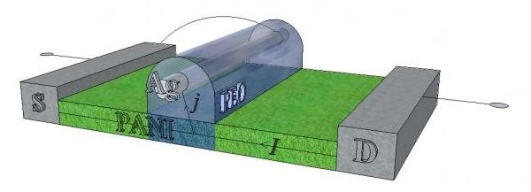
A group of scientists has created a neural network based on polymeric memristors — devices that can potentially be used to build fundamentally new computers. These developments will primarily help in creating technologies for machine vision, hearing, and other machine sensory systems, and also for intelligent control systems in various fields of applications, including autonomous robots.

Read more

An international team of researchers has developed a new algorithm that could one day help scientists reprogram cells to plug any kind of gap in the human body. The computer code model, called Mogrify, is designed to make the process of creating pluripotent stem cells much quicker and more straightforward than ever before.

A pluripotent stem cell is one that has the potential to become any type of specialised cell in the body: eye tissue, or a neural cell, or cells to build a heart. In theory, that would open up the potential for doctors to regrow limbs, make organs to order, and patch up the human body in all kinds of ways that aren’t currently possible.

It was Japanese researcher Shinya Yamanaka who first reprogrammed cells in this way back in 2007 — it later earned him a Nobel Prize — but Yamanaka’s work involved a lot of labourious trial and error, and the process he followed is not an easy one to reproduce. Mogrify aims to compute the required set of factors to change cells instead, and it’s passed its early tests with flying colours.

Read more

As recently as 50 years ago, psychiatry lacked a scientific foundation, the medical community considered mental illness a disorder of the mind, and mental patients were literally written off as “sick in the head.” A fortunate turn in progress has yielded today’s modern imaging devices, which allow neuroscientists and psychiatrists to examine the brain of an individual suffering from a mental disorder and provide the best treatment options. In a recent interview, Columbia University Psychiatry Chair Dr. Jeffrey Lieberman stated that new research into understanding the mind is growing at an accelerated pace.

(iStock)
(iStock)

Lieberman noted that, just as Galileo couldn’t prove heliocentrism until he had a telescope, psychiatry lacked the technological sophistication, tools, and instruments necessary to get an understanding of the brain until the 1950s. It wasn’t until the advent of psychopharmacology and neuroimaging, he said, that researchers could look inside the so-called black box that is the brain.

“(It began with) the CAT scan, magnetic resonance imaging (MRI) systems, positron emission tomography (PET scans) and then molecular genetics. Most recently, the burgeoning discipline of neuroscience and all of the methods within, beginning with molecular biology and progressing to optogenetics, this capacity has given researchers the ability to deconstruct the brain, understand its integral components, its mechanisms of action and how they underpin mental function and behavior,” Lieberman said. “The momentum that has built is almost like Moore’s law with computer chips, (and) you see this increasing power occurring with exponential sort of growth.”

Specifically, the use of MRIs and PET scans has allowed researchers to study the actual functional activity of different circuits and regions of the brain, Lieberman noted. Further, PET scans provided a look at the chemistry of the brain, which has allowed for the development of more sophisticated pathological theories. These measures, he said, were used to develop treatments while also allowing measurement of the effectiveness of both medication-based therapies and psychotherapies.

As an example, Lieberman cited the use of imaging in the treatment of post-traumatic stress disorder (PTSD). The disorder, a hyperarousal that chronically persists even in the absence of threatening stimulation, is treated through a method called desensitization. Over time, researchers have been able to fine-tune the desensitization therapies and treatments by accessing electronic images of the brain, which can show if there’s been a reduction in the activation of the affected amygdala.

Lieberman noted that despite progress in this area, technology has not replaced interaction with the individual patient; however, as technology continues to evolve, he expects the diagnoses of mental disorders to be refined.

“By the use of different technologies including genetics (and) imaging, including electrophysiological assessments, which are kind of EEG based, what we’ll have is one test that can confirm conditions that were previously defined by clinical description of systems,” Lieberman said. “I think, of all the disciplines that will do this, genetics will be the most informative.”

Just as genetics is currently used to diagnose cancer using anatomy and histology, Lieberman said the expanding field is helping researchers distinguish mental illness in individuals with certain genetic mutations. He expects that in the future, doctors will use “biochips” to routinely screen patients and provide a targeted therapy against the gene or gene product. These chips will have panels of genes known to be potentially associated with the risk for mental illness.

“Someone used the analogy of saying the way we treat depression now is as if you needed to put coolant into your car. Instead of putting it into the radiator, you just dump it on the engine,” he said. “So genetics will probably be the most powerful method to really tailor to the individual and use this technique of precision and personalized medicine.”

Lieberman also sees additional promise in magnetic stimulation, deep brain stimulation through the surgical implanting of electrodes, and optogenetics. Though he has plenty of optimism for these treatments and other potential treatments for mental illness, much of their continued growth may hinge on government policy and budgets. Recent coverage of gun violence in the United States, and a public call for better means by which to screen individuals for mental health inflictions, may be an unfortunate catalyst in moving funding forward in this research arena. A recent article from the UK’s Telegraph discusses Google’s newfound interest in this research, with former US Head of the National Institute of Mental Health now in a position at Google Life Sciences.

“Science, technology and healthcare are doing very well, but when it comes to the governmental process, I think we’re in trouble,” he said. “A welcome development in this regard is President Obama’s Human Brain Initiative, which if you look at the description of it, (is) basically to develop new tools in neurotechnology that can really move forward in a powerful way of being able to measure the function of the brain. Not by single cells or single circuits, but by thousands or tens of thousands of cells and multiple circuits simultaneously. That’s what we need.”

Luv the whole beautiful picture of a Big Data Quantum Computing Cloud. And, we’re definitely going to need it for all of our data demands and performance demands when you layer in the future of AI (including robotics), wearables, our ongoing convergence to singularity with nanobots and other BMI technologies. Why we could easily exceed $4.6 bil by 2021.


From gene mapping to space exploration, humanity continues to generate ever-larger sets of data—far more information than people can actually process, manage, or understand.

Machine learning systems can help researchers deal with this ever-growing flood of information. Some of the most powerful of these analytical tools are based on a strange branch of geometry called topology, which deals with properties that stay the same even when something is bent and stretched every which way.

Such topological systems are especially useful for analyzing the connections in complex networks, such as the internal wiring of the brain, the U.S. power grid, or the global interconnections of the Internet. But even with the most powerful modern supercomputers, such problems remain daunting and impractical to solve. Now, a new approach that would use quantum computers to streamline these problems has been developed by researchers at MIT, the University of Waterloo, and the University of Southern California…

More insights for AI research.


The complex multi-stage architecture of cortical visual pathways provides the neural basis for efficient visual object recognition in humans. However, the stage-wise computations therein remain poorly understood. Here, we compared temporal (magnetoencephalography) and spatial (functional MRI) visual brain representations with representations in an artificial deep neural network (DNN) tuned to the statistics of real-world visual recognition. We showed that the DNN captured the stages of human visual processing in both time and space from early visual areas towards the dorsal and ventral streams. Further investigation of crucial DNN parameters revealed that while model architecture was important, training on real-world categorization was necessary to enforce spatio-temporal hierarchical relationships with the brain. Together our results provide an algorithmically informed view on the spatio-temporal dynamics of visual object recognition in the human visual brain.

Read more