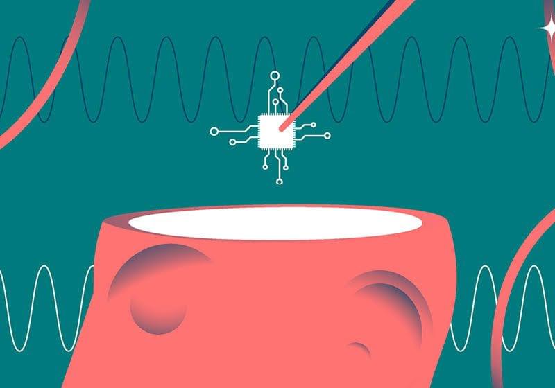
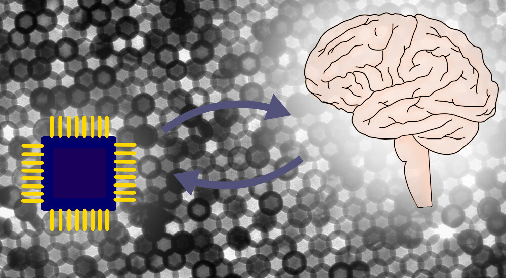
Microscopic wireless electronic devices hitch a ride on immune cells to reach the brain, offering therapeutic avenues for brain cancers and neurodegeneration.

Patterns of smartphone use and their impact on mental health are being extensively studied due to the growing dependence of the device in people’s lives.
A recent study tracked late-night smartphone usage in 79 adults with recent suicidal thoughts for 28 days. People using phones late between 11 p.m. and 1 a.m. showed a higher risk of suicidal thoughts the next day, whereas those who actively used the keyboard beyond midnight hours showed a lower risk.
The findings are published in JAMA Network Open.


Interfacing artificial devices with the human brain is the central goal of neurotechnology. Yet, our imaginations are often limited by currently available paradigms and technologies. Suggestions for brain–machine interfaces have changed over time, along with the available technology. Mechanical levers and cable winches were used to move parts of the brain during the mechanical age. Sophisticated electronic wiring and remote control have arisen during the electronic age, ultimately leading to plug-and-play computer interfaces. Nonetheless, our brains are so complex that these visions, until recently, largely remained unreachable dreams. The general problem, thus far, is that most of our technology is mechanically and/or electrically engineered, whereas the brain is a living, dynamic entity. As a result, these worlds are difficult to interface with one another.
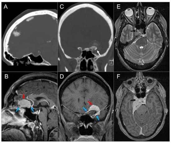
The skull base provides a platform for supporting the brain while serving as a conduit for major neurovascular structures. In addition to malignant lesions originating in the skull base, there are many benign entities and developmental variants that may simulate disease. Therefore, a basic understanding of the relevant embryology is essential. Lesions centered in the skull base can extend to the adjacent intracranial and extracranial compartments; conversely, the skull base can be secondarily involved by primary extracranial and intracranial disease. CT and MRI are the mainstay imaging methods and are complementary in the evaluation of skull base lesions. Advances in cross-sectional imaging have been crucial in the management of patients with skull base pathology, as this represents a complex anatomical area that is hidden from direct clinical exam.
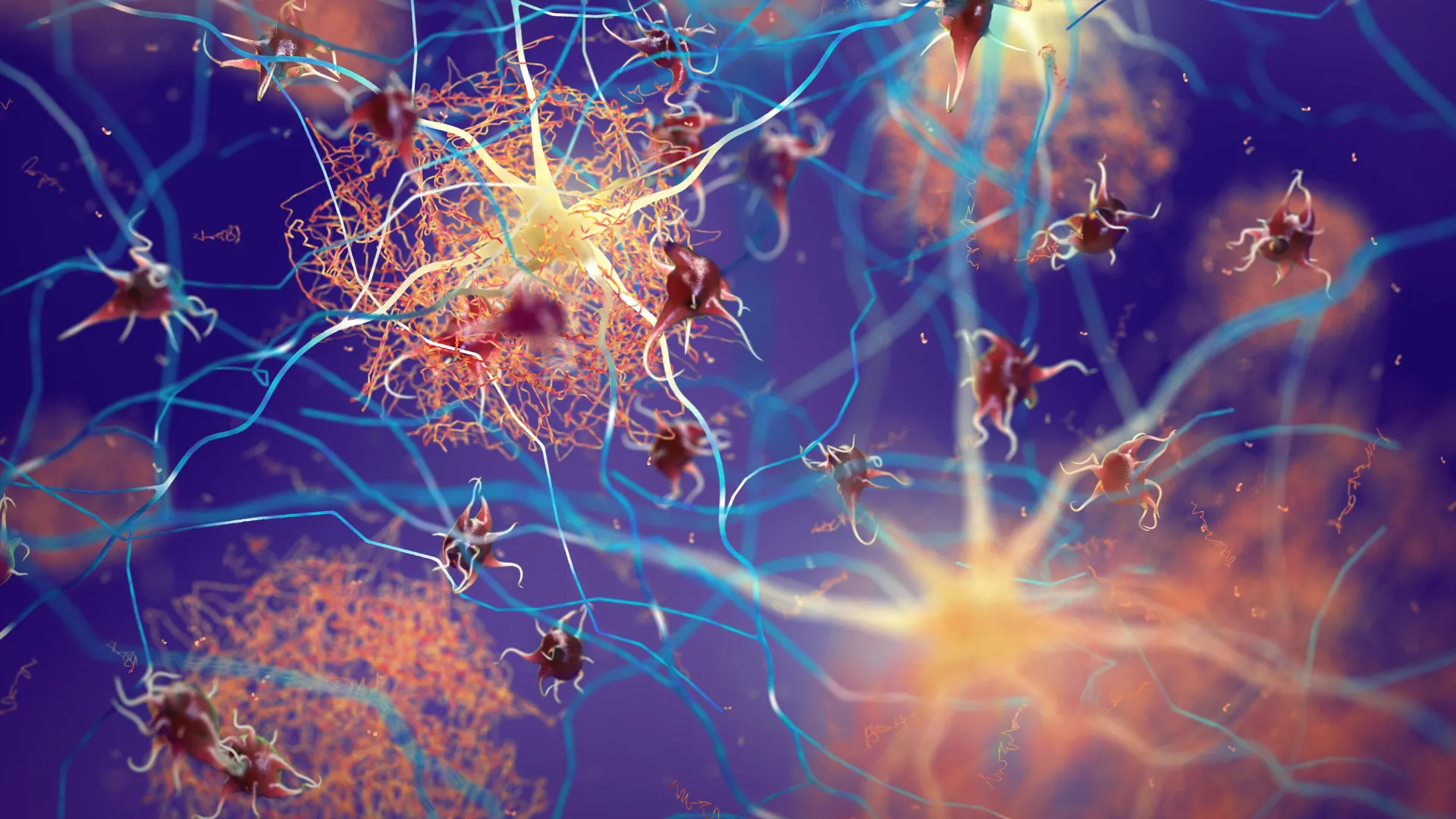
Scientists have developed a new molecule that breaks down beta-amyloid plaques by binding to excess copper in the brain. The treatment restored memory and reduced inflammation in rats, while also proving non-toxic and able to cross the blood–brain barrier. Because it’s far simpler and potentially cheaper than existing drugs, researchers are now pursuing partnerships to begin human trials.

The ratio of two dominant groups of microbes in the human gut was higher across all three disorder groups than was typically seen in the control group.
A new, small study suggests children with autism, ADHD, and anorexia share similarly disrupted gut microbiomes, which, by some measures, have more in common with each other than with their healthy, neurotypical peers.
Led by researchers from Comenius University in Slovakia, the study used stool samples to assess the gut microbiomes of 117 children.
The exploratory study included 30 boys with autism spectrum disorder (ASD), 21 girls with anorexia nervosa, and 14 children with attention deficit hyperactivity disorder (ADHD). The remaining samples were from age-and sex-matched healthy and neurotypical children, providing a control group.

Researchers from Curtin University in Australia and multiple universities in Ethiopia report that prenatal folic acid and multivitamin supplementation is associated with a roughly 30% lower risk of autism spectrum disorder (ASD) in children, based on an umbrella review of existing systematic reviews and meta-analyses.
Global estimates in the reviewed material place ASD prevalence at up to 1% of children. ASD affects reciprocal social interaction, nonverbal communication, and understanding of social relationships. Co-occurring conditions frequently include epilepsy, depression, anxiety, attention deficit hyperactivity disorder, sleep disturbance, and self-injury.
Previous studies found that both genetic mutations and environmental influences contribute to ASD risk, with prenatal maternal nutrition identified as one modifiable environmental factor. Within that broader category of prenatal maternal nutrition, folic acid and multivitamin supplements are among the most accessible interventions offered to women before and during pregnancy.

Temporarily anesthetizing the retina briefly reverts the activity of the visual system to that observed in early development and enables growth of responses to the amblyopic eye, new research shows.
In the common vision disorder amblyopia, impaired vision in one eye during development causes neural connections in the brain’s visual system to shift toward supporting the other eye, leaving the amblyopic eye less capable even after the original impairment is corrected. Current interventions are only effective during infancy and early childhood while the neural connections are still being formed.
But a new study in mice by neuroscientists in The Picower Institute for Learning and Memory at MIT shows that if the retina of the amblyopic eye is temporarily and reversibly anesthetized just for a couple of days, the brain’s visual response to the eye can be restored even in adulthood.