Toggle light / dark theme

Galimedix starts phase 2 fundraise after oral Alzheimer’s candidate clears safety test

Maryland-based biotech Galimedix Therapeutics is ready to take a big swing on its oral Alzheimer’s disease candidate after no serious adverse events were reported and the small molecule successfully crossed the blood-brain barrier in a phase 1 trial.

The first-ever clinical trial for GAL-101 tested single and multiple ascending doses of the asset in more than 100 healthy volunteers, Galimedix reported in a Sept. 12 release. The company is now initiating fundraising for a phase 2 trial.

Mini microscope enables real-time 3D brain imaging in freely moving mice

Researchers at the University of California, Davis, have created a miniaturized microscope for real-time, high-resolution, noninvasive imaging of brain activity in mice. The device is a significant step toward revolutionizing how neuroscientists study the brain.

“What we are doing is creating technology to image brain activity in freely moving and behaving mice to open up the behavior paradigm,” said Weijian Yang, professor of electrical and computer engineering. “The goal is to create a device capable of enabling research into brain activity and behavior in mice in real time—to see how brain activity drives behavior or perception.”

The microscope will advance insights into how the brain works, which is expected to benefit human health by empowering the development of new and improved therapeutic strategies for brain disorders.

The origin of the mental number line may be biological, not cultural, according to a new study

A new study has found that a chick’s ability to mentally organize numbers along a line from left to right is not learned but is instead a direct result of brain specialization that occurs before hatching. Researchers found that exposing chick eggs to light in the final days of incubation causes the two hemispheres of the brain to develop distinct functions, which in turn establishes an innate tendency for the chicks to count from left to right.

For many years, scientists and philosophers have debated the origins of the “mental number line,” a common intuition where people visualize smaller numbers on the left and larger numbers on the right. The prevailing theory suggested this was a cultural artifact, learned through years of reading and writing in a left-to-right direction.

However, this idea has been challenged by findings that pre-verbal infants and even some animals exhibit a similar spatial bias for numbers, suggesting a deeper, biological foundation. Researchers have hypothesized that this foundation lies in brain lateralization, the process where the left and right hemispheres of the brain become specialized for different cognitive tasks. While this connection seemed plausible, there was little direct experimental evidence to confirm that brain specialization actually causes this numerical mapping.

Clinical Trial To Transform Alzheimer’s Diagnosis Launched in UK

UK researchers on Wednesday announced the trial of a blood test for Alzheimer’s, which it is hoped will transform the diagnosis of the disease.

Researchers at University College London (UCL) will assess whether the test could improve the accuracy of diagnosis from 70 percent to over 90 percent.

Medics say early diagnosis is critical with Alzheimer’s, which is the most common cause of dementia, as the earlier treatment is started, the more effective it is.

New study links blood proteins to Alzheimer’s disease and memory loss

Researchers at Emory Goizueta Brain Health Institute and partner institutions have found new clues in the blood that could help explain why Alzheimer’s disease develops and how it affects memory.

The study, published in Nature Aging, examined from more than 2,100 individuals across four large research cohorts. Using advanced tools, scientists measured thousands of proteins in the blood and linked them to changes in the and thinking ability.

Traditionally, doctors have focused on sticky amyloid plaques in the brain as a hallmark of Alzheimer’s.

Genetic deletion in cerebellum impedes hemisphere formation, study finds

The cerebellum, a brain region located at the back of the head that has long been known to support the coordination of muscle movements, has recently also been implicated in more sophisticated mental functions. Purkinje cells are the only neurons located in the cerebellum that integrate information in the cerebellar cortex and send it to other parts of the nervous system.

Purkinje cells are large and highly branched that can have different functions. While many past studies have explored the roles of these cells, the neural and genetic processes shaping their diversity have not yet been fully elucidated.

Researchers at the University of Connecticut School of Medicine recently carried out a study aimed at exploring the possible role of the FOXP genes, a family of genes known to contribute to switching other genes “on and off,” in shaping Purkinje cell populations and the formation of circuits in the . Their findings, published in Nature Neuroscience, hint at the existence of at least 11 different Purkinje cell subtypes, suggesting that the FOXP1 and FOXP2 genes contribute to their diversification.

Soft magnetoelastic sensor measures fatigue from eyeball movements in real-time

Over the past few decades, electronics engineers have developed increasingly sophisticated sensors that can reliably measure a wide range of physiological signals, including heart rate, blood pressure, respiration rate and oxygen saturation. These sensors were used to create both biomedical and consumer-facing wearable devices, advancing research and the real-time monitoring of health-related metrics, such as sleep quality and physiological stress.

Fatigue, a mental state marked by a decline in performance due to stress, lack of sleep, excessive activity or other factors, has proved to be more difficult to reliably quantify. Most existing methods for measuring fatigue rely on surveys that ask people to report how tired they feel, a method to record the brain’s electrical activity known as electroencephalography (EEG) or camera-based systems.

Most of these approaches are unreliable or only applicable in laboratory settings, as they rely on subjective evaluations, bulky equipment or controlled environments. These limitations prevent their large-scale deployment in everyday settings.

‘Young’ immune cells reverse signs of neurodegenerative brain changes in preclinical study

“Young” immune cells created by Cedars-Sinai investigators reversed signs of aging and Alzheimer’s disease in the brains of laboratory mice, according to a study published in the journal Advanced Science.

The immune cells, which were produced from human stem cells, could be used to develop new treatments for neurological conditions in humans.

“Previous studies have shown that transfusions of blood or plasma from young mice improved cognitive decline in older mice, but that is difficult to translate into a therapy,” said Clive Svendsen, Ph.D., executive director of the Board of Governors Regenerative Medicine Institute and senior author of the study.

Personalized brain stimulation shows benefit for depression

A more precise and personalized form of electric brain stimulation may be a more effective and faster treatment for people with moderate to major depression compared to other similar treatments, according to a UCLA Health study.

The study, published in JAMA Network Open, examined the effectiveness of a noninvasive brain stimulation treatment known as (HD-tDCS) in treating depression. Transcranial direct current stimulation uses electrodes placed on a patient’s scalp to deliver noninvasive, safe levels of electrical currents to targeted areas of the brain.

For depression, the treatment is used to target brain networks that regulate emotional processing and self-referential thoughts. TDCS has not been approved by the U.S. Food and Drug Administration as a treatment for depression, and into various forms of tDCS is ongoing.

Distinct psilocybin-induced oscillations observed in rat medial prefrontal cortex, with effects lasting days

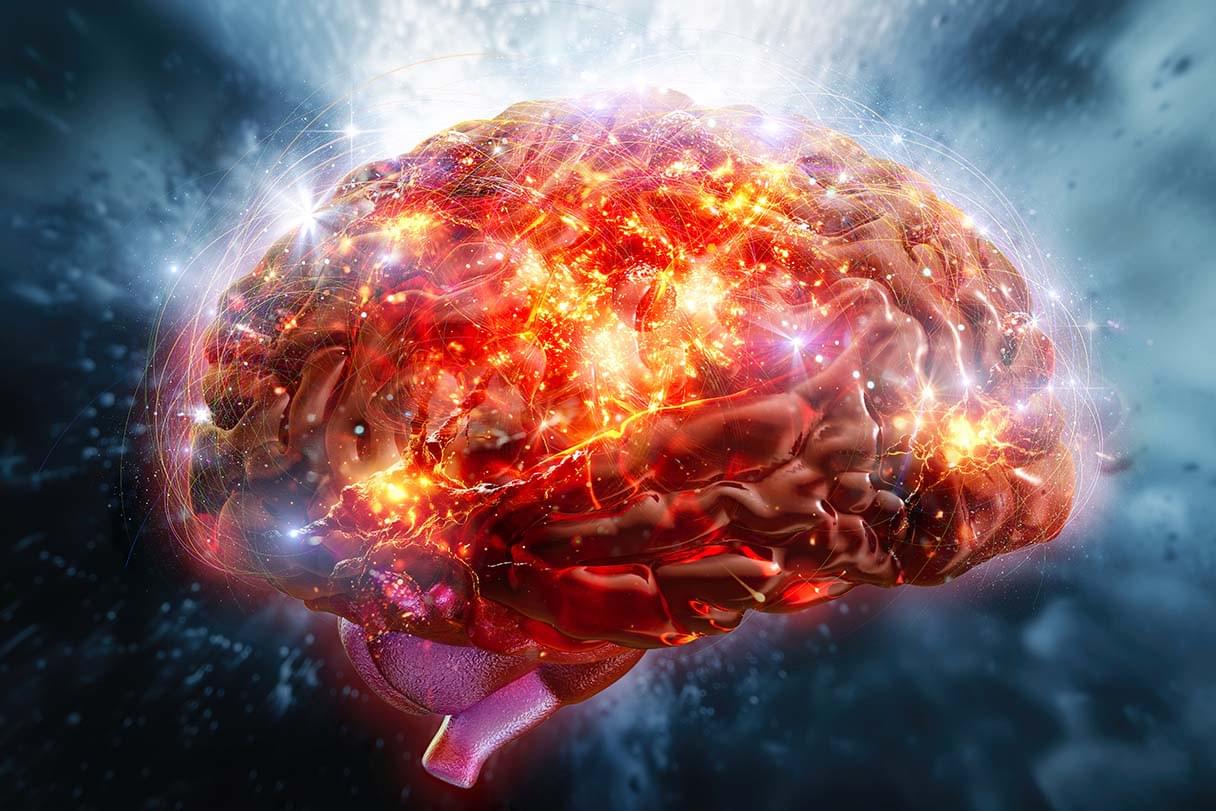
Psychedelics, a class of psychoactive drugs that typically induce peculiar mental states and hallucinations, are still prohibited for recreational use in most countries worldwide. In recent years, some neuroscientists and medical researchers have been exploring the potential therapeutic effects of these drugs, focusing on the treatment of depression, anxiety and various substance use disorders.

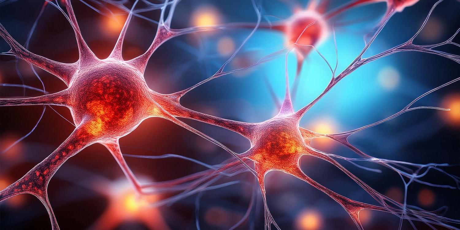
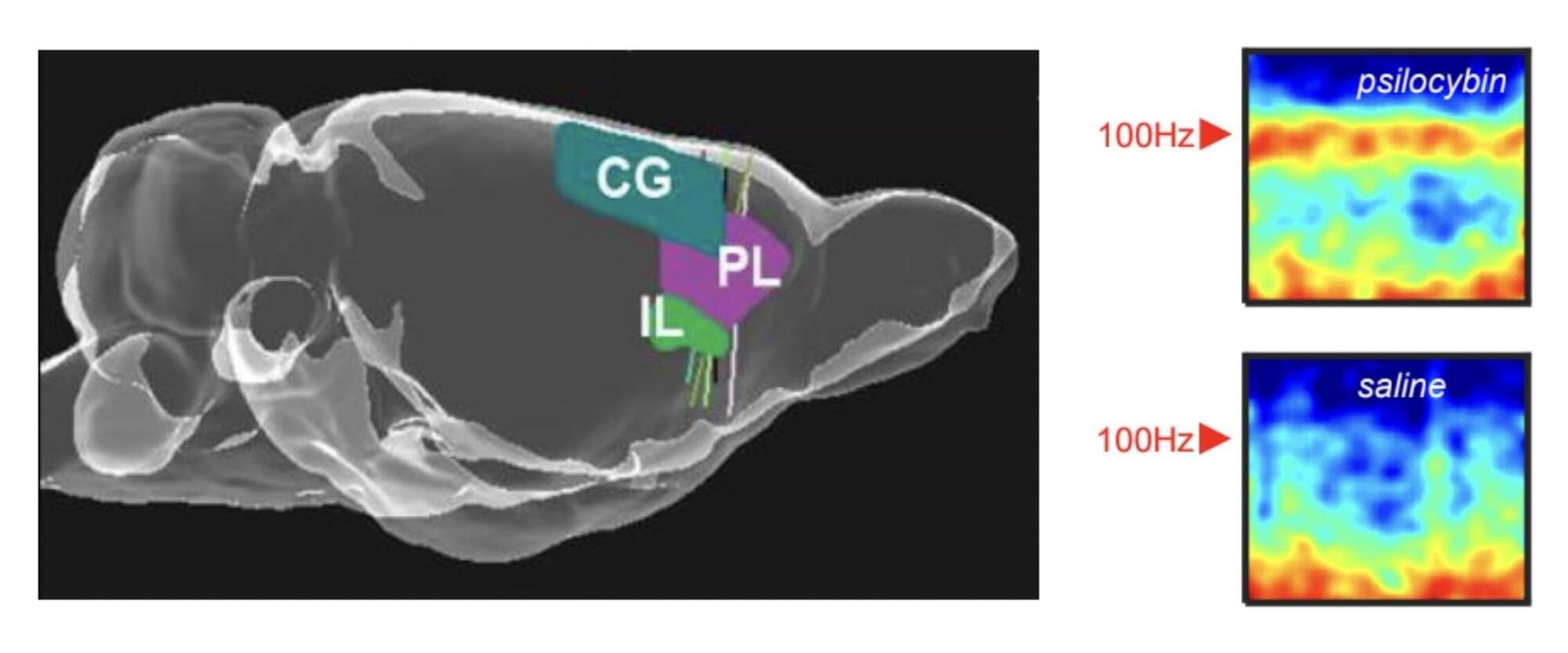
Researchers at the University of Bristol, Compass Pathways plc and other institutes recently carried out a new study involving rats, exploring the effects of the psychedelic compound on the activity of neurons in the medial prefrontal cortex, a brain region that supports decision-making, attention and the regulation of emotions. Their paper, published in Molecular Psychiatry, outlines some of the associated with the intake of this compound, which had not yet been observed in human experiments.

“Psychedelic drugs like have profound effects on our brains and minds,” Matt Jones, Professor of Neuroscience at the University of Bristol and senior author of the paper, told Medical Xpress. “These effects are fascinating and—as a long history of psychedelic use and recent clinical trials attest—potentially beneficial. This study was driven by two interrelated questions. Firstly, how does a relatively simple, small molecule alter brain activity to completely change our mental model of the world? Secondly, can those effects be harnessed to help treat mental illness?”

/* */