A single indulgent meal may carry hidden risks. Scientists found that drinking a milkshake with 130 grams of fat impaired blood flow to the brain within hours, raising concerns about stroke, dementia, and how everyday diets shape brain health.


The anterior insular cortex (aIC) is an important brain region known to contribute to the regulation of emotions, the integration of bodily sensations, decision-making and some other functions. Past studies have linked this brain region to some neuropsychiatric disorders characterized by unusual patterns of thinking and behavior, including autism spectrum disorder (ASD) and depression.
However, the precise cellular and neurobiological processes via which the aIC might contribute to ASD and depression have not yet been clearly elucidated. Some neuroscientists have been exploring the possibility that microglia, immune cells that play a role in eliminating damaged cells and pathogens, could play a role in some of the behaviors linked with these two neuropsychiatric disorders.
Researchers at Tsinghua University recently carried out a study involving mice, aimed at investigating the possibility that microglia in the aIC play a part in some of the symptoms of ASD and depression. Their paper, published in Molecular Psychiatry, identifies two distinct subtypes of microglia that appear to contribute to autism-like and depression-like behavior in mice.
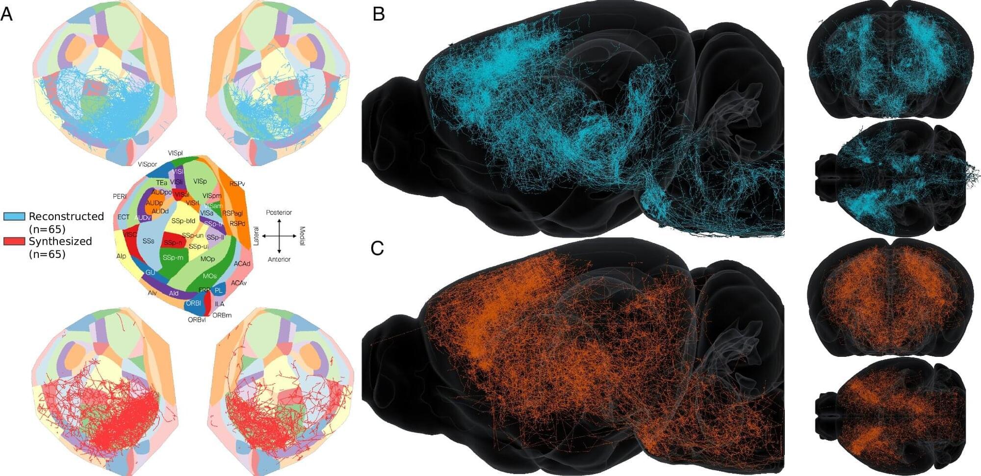
EPFL researchers have developed a powerful method to generate brain-wide, biologically realistic wiring maps of the mouse brain. Their approach bridges experimental data with mathematical and computational modeling to simulate how neurons connect across the entire brain.
The study is published in the journal Nature Communications.
One of neuroscience’s greatest challenges is understanding how the brain is wired. Even with modern imaging tools, it has been a challenge to create detailed maps that show how the brain’s billions of cells (neurons) connect, not just with their local “neighbors” but also to other, more distant cells in the brain.

One of the defining features of humans is our brain’s remarkable capacity for language, planning, memory, creativity, and more. These abilities stem not just from our large brain size, but also from the folded structure of the brain’s outer layer, the cerebral cortex.
A new study, published in the journal Nature Communications, offers insight into how these wrinkles form, pointing to a range of contributing factors—including the number of early-stage brain cells, how they migrate during development, and the specific types of cells involved.
These findings may help guide future research into brain development, evolution, and health.


By boosting the activity of cellular ‘power stations’ in the brains of mice with a dementia-like condition, an international team of researchers has reversed pathological memory loss.
Problems with energy-producing cellular structures called mitochondria have previously been linked to neurodegenerative diseases such as Alzheimer’s. Before now, it wasn’t clear if this was a cause or a consequence of these conditions.
“This work is the first to establish a cause-and-effect link between mitochondrial dysfunction and symptoms related to neurodegenerative diseases, suggesting that impaired mitochondrial activity could be at the origin of the onset of neuronal degeneration,” says Giovanni Marsicano, a neuroscientist from the French National Institute of Health and Medical Research (INSERM).
