Toggle light / dark theme

Scientists discover how to reactivate cancer’s molecular “kill switch”

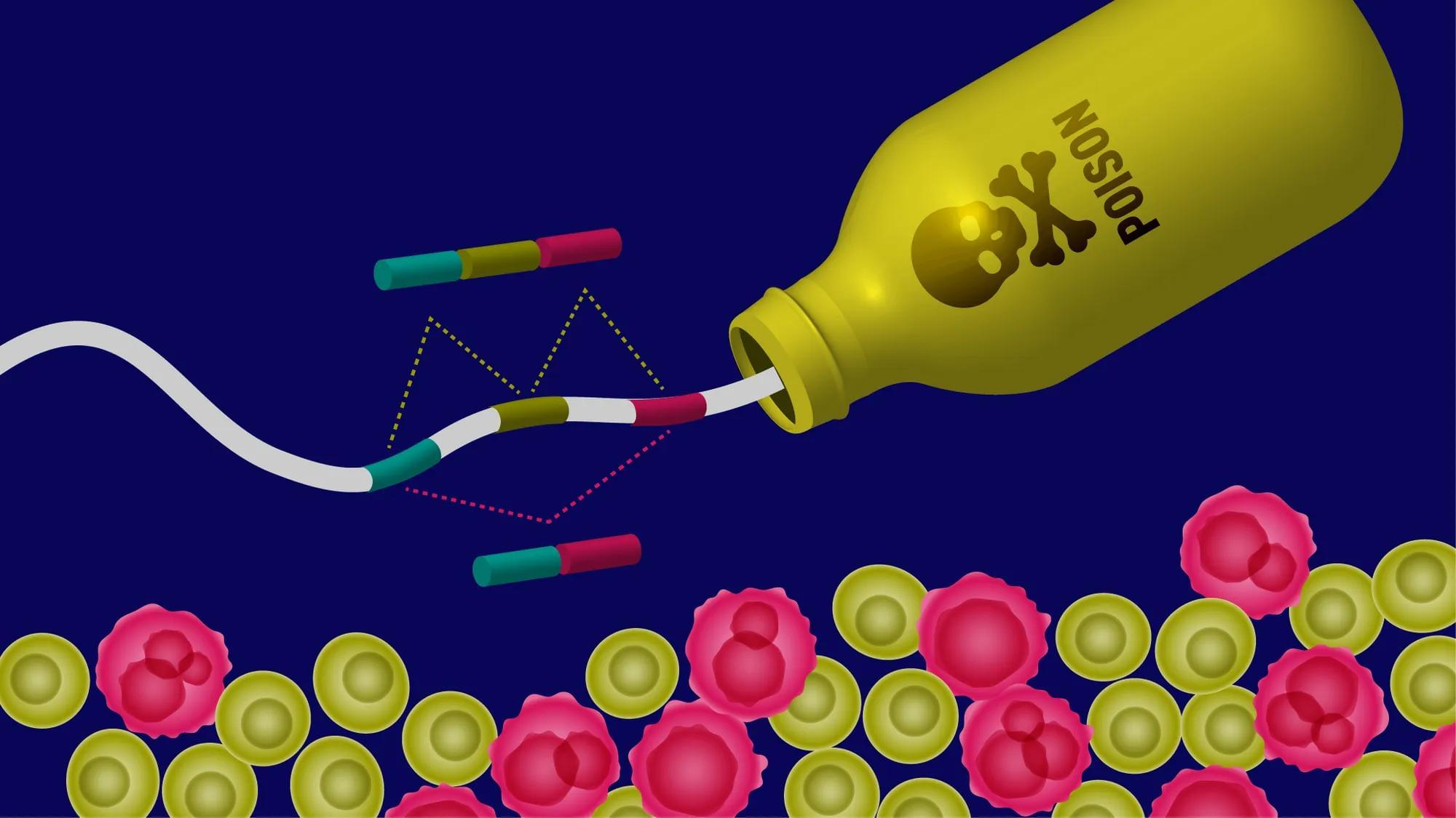
(Farmington, Conn. – March 13, 2025) – Alternative RNA splicing is like a movie editor cutting and rearranging scenes from the same footage to create different versions of a film. By selecting which scenes to keep and which to leave out, the editor can produce a drama, a comedy, or even a thriller—all from the same raw material. Similarly, cells splice RNA in different ways to produce a variety of proteins from a single gene, fine-tuning their function based on need. However, when cancer rewrites the script, this process goes awry, fueling tumor growth and survival.

In a recent study reported in the Feb. 15 issue of Nature Communications, scientists from The Jackson Laboratory (JAX) and UConn Health not only show how cancer hijacks this tightly regulated splicing and rearranging of RNA but also introduce a potential therapeutic strategy that could slow or even shrink aggressive and hard-to-treat tumors. This discovery could transform how we treat aggressive cancers like triple-negative breast cancer and certain brain tumors, where current treatment options are limited.

At the heart of this work, led by Olga Anczuków, an associate professor at JAX and co-program leader at the NCI-designated JAX Cancer Center, are tiny genetic elements called poison exons, nature’s own “off switch” for protein production. When these exons are included in an RNA message, they trigger its destruction before a protein can be made—preventing harmful cellular activity. In healthy cells, poison exons regulate the levels of key proteins, keeping the genetic machinery in check. But in cancer, this safety mechanism often fails.

Genetic identification of mouse trigeminal afferents responsible for mechanical allodynia

Peripheral mechanisms by which the nerve afferents send signals after nerve injury in mechanical C is not well understood.

The researchers identify a subset of trigeminal afferents in mice activated by facial brushing after nerve injury. Many of brushing-activated (BA) trigeminal ganglia (TG) neurons express neurofilament200 and Ntrk3, markers for low-threshold mechanoreceptors, with lower co-localization with nociceptor markers such as Calca or Trpv1.

Inhibition of BA TG neurons reduces mechanical allodynia, while their activation increases spontaneous face wiping after nerve injury.

Knockdown of Piezo2 from BA TG afferents reduces mechanical allodynia. Thus, targeted manipulations reveal these neurons’ causal role in pain-like behaviors and heightened sensitivity, providing new insights into primary afferents underlying mechanical allodynia. sciencenewshighlights ScienceMission https://sciencemission.com/trigeminal-afferents


Peripheral mechanisms of mechanical allodynia after nerve injury remain unclear. Li et al. identify a subset of trigeminal afferents in mice activated by facial brushing after nerve injury. Targeted manipulations reveal these neurons’ causal role in pain-like behaviors and heightened sensitivity, providing new insights into primary afferents underlying mechanical allodynia.

A process thought to destroy brain cells might actually help them store data

To ensure these laboratory-created fibers were the same as those found in living brains, the team utilized cryogenic electron microscopy (cryo-EM). This advanced imaging technique allows scientists to see the atomic structure of proteins.

The images revealed that the Orb2 amyloids created with the help of Funes were structurally identical to endogenous Orb2 amyloids extracted from fly heads. They possessed the same “cross-beta” architecture that characterizes functional amyloids.

The study further demonstrated that the “J-domain” of the Funes protein is essential for this activity. This domain is a specific section of the protein sequence that defines the JDP family.

Missing Link Between Parkinson’s Protein And Damage to Brain Cells Discovered

An investigation by researchers from Case Western Reserve University School of Medicine in the US has filled in a missing link between the toxic build-up of proteins in the neurodegenerative condition Parkinson’s disease and the death of critical brain cells.

The result of three years of research, the discovery connects alpha-synuclein proteins to a breakdown in mitochondrial function, both previously linked to Parkinson’s.

“We’ve uncovered a harmful interaction between proteins that damages the brain’s cellular powerhouses, called mitochondria,” says neuroscientist Xin Qi.

Long-term antidepressant effects of psilocybin linked to functional brain changes

In the group treated with psilocybin, adapting neurons sat at a resting voltage that was closer to the threshold for firing. This state is known as depolarization. It means the cells are primed to activate more easily. The bursting neurons in psilocybin-treated rats also showed increased excitability. They required less input to trigger a signal and fired at faster rates than neurons in untreated rats.

The rats treated with 25CN-NBOH also exhibited functional changes, though the specific electrical alterations differed slightly from the psilocybin group. For instance, the bursting neurons in this group were not as easily triggered as those in the psilocybin group. However, the overall pattern confirmed that the drug had induced a lasting shift in neuronal function.

These electrophysiological findings provide a potential explanation for the behavioral results. While the physical branches of the neurons may have pruned back to normal levels, the cells “remembered” the treatment through altered electrical tuning. This functional shift allows the neural circuits to operate differently long after the drug has left the body.

CRISPR screens in iPSC-derived neurons reveal principles of tau proteostasis

Now online! CRISPR screens in iPSC-derived neurons reveal that the E3 ubiquitin ligase CRL5SOCS4 ubiquitinates tau, that CUL5 expression is correlated with resilience in human Alzheimer’s disease, and that electron transport chain dysfunction alters tau degradation by the proteasome.

Spaceflight causes astronauts’ brains to shift, stretch and compress in microgravity

Spaceflight takes a physical toll on astronauts, causing muscles to atrophy, bones to thin and bodily fluids to shift. According to a new study published in the journal PNAS, we can now add another major change to that list. Being in microgravity causes the brain to change shape.

Here on Earth, gravity helps to keep the brain anchored in place while the cerebrospinal fluid that surrounds it acts as a cushion. Scientists already knew that, without gravity’s steady pull, the brain moves upward, but this new research showed that it is also stretched and compressed in several areas.

Brains on the move Researchers led by Rachel Seidler at the University of Florida reached this conclusion after studying MRI scans of 26 astronauts taken before and after their missions to the International Space Station. These were compared with scans from 24 volunteers who participated in a head-down tilt bed rest experiment. They spent 60 days lying at a six-degree downward angle to mimic how weightlessness causes bodily fluids and organs to move toward the head.

/* */