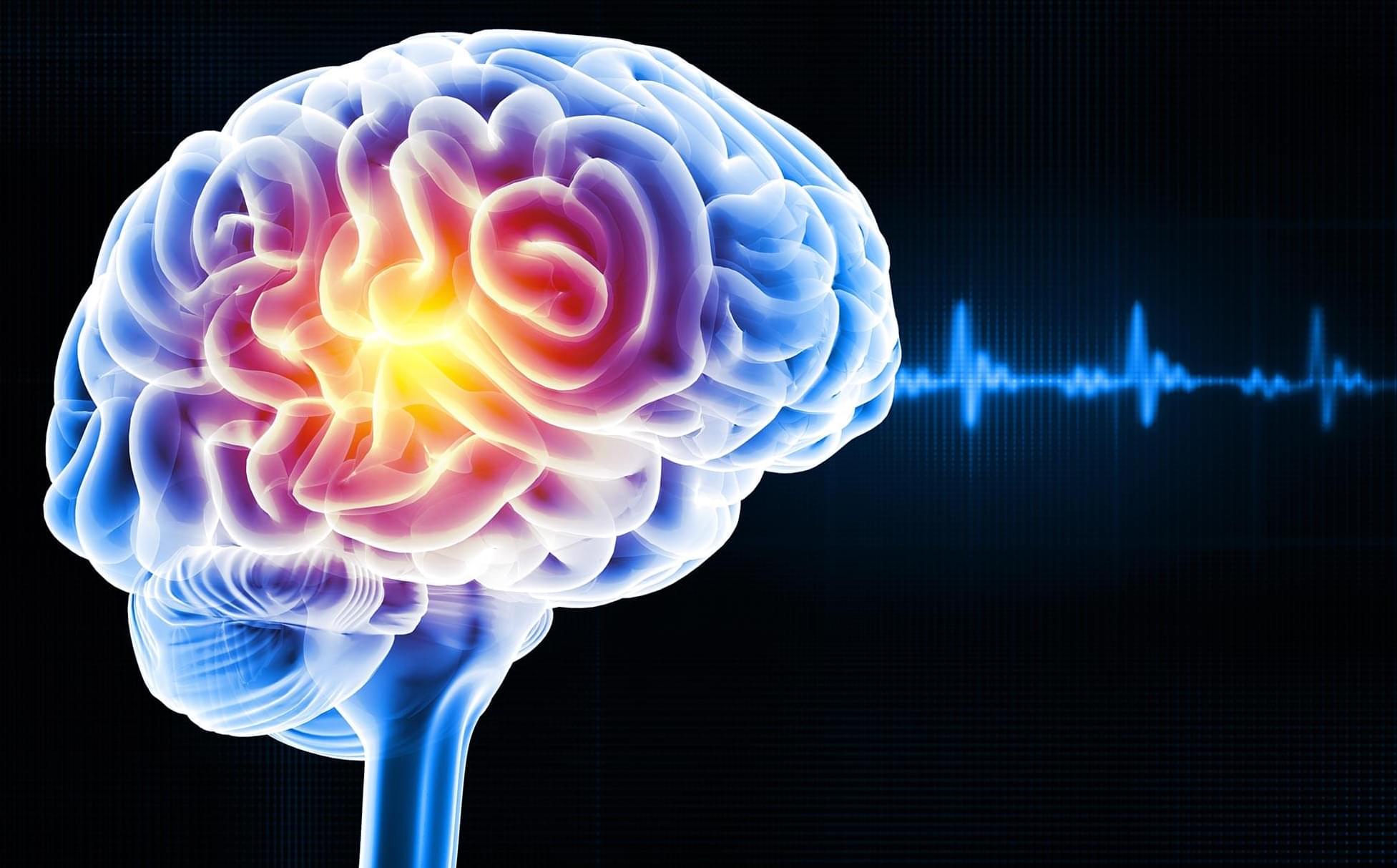
Toggle light / dark theme

How sleep microstructure organizes memory replay.

T occur in isolation; older memories are also replayed during sleep, raising an intriguing challenge: how does the brain avoid interference between fragile new memories and stable old ones?” + To explore this question, researchers developed a groundbreaking method to study both hippocampal activity and sleep dynamics simultaneously in naturally sleeping mice. Using a technique called pupillometry, which measures oscillatory changes in pupil size, they uncovered a previously unknown “microstructure” within non-REM sleep that helps the brain manage memory replay.

They discovered that memory replay is organized into distinct substates of non-REM sleep:

1. Contracted pupil substates: During these phases, the hippocampus predominantly replays new memories. This activity is associated with sharp-wave ripples—brief bursts of electrical activity critical for memory consolidation—and strong excitatory inputs from external sources.

2. Dilated pupil substates: In contrast, older memories are reactivated during these phases, characterized by increased local inhibitory activity, which helps maintain stability and prevent interference.

Research reveals distinct mechanisms underlying neonatal and post-pubertal social behaviors, providing valuable insights for developing targeted early interventions.

Researchers from the University of Texas Health Science Center at San Antonio and Hirosaki University have unveiled significant findings on the development of social behaviors in fragile X syndrome, the most common genetic cause of autism spectrum disorder. The study, published in Genomic Psychiatry, highlights the effects of a specific prenatal treatment on social behaviors in mice.

The researchers found that administering bumetanide—a drug that regulates chloride levels in neurons—to pregnant mice restored normal social communication in newborn pups with the fragile X mutation. However, they also discovered an unexpected outcome: the same treatment reduced social interaction after puberty in both fragile X and typical mice. These findings shed light on the complex and developmental-stage-specific effects of interventions for fragile X syndrome.

Largest brain aging study points to possible connections between diet, inflammation, and brain health.

Scientists at the Allen Institute have discovered specific types of brain cells in mice that experience significant changes as they age. They also identified a distinct “hotspot” where many of these changes are concentrated. Published today (January 1) in Nature, these findings could lead to the development of therapies aimed at slowing or managing the brain’s aging process.

Sensitive cells: Scientists discovered dozens of specific cell types, mostly glial cells, known as brain support cells, that underwent significant gene expression changes with age. Those strongly affected included microglia and border-associated macrophages, oligodendrocytes, tanycytes, and ependymal cells.

Inflammation and neuron protection: In aging brains, genes associated with inflammation increased in activity while those related to neuronal structure and function decreased.

Aging hot spot: Scientists discovered a specific hot spot combining both the decrease in neuronal function and the increase in inflammation in the hypothalamus. The most significant gene expression changes were found in cell types near the third ventricle of the hypothalamus, including tanycytes, ependymal cells, and neurons known for their role in food intake, energy homeostasis, metabolism, and how our bodies use nutrients. This points to a possible connection between diet, lifestyle factors, brain aging, and changes that can influence our susceptibility to age-related brain disorders.

Brain-wide cell-type-specific transcriptomic signatures of healthy ageing in mice.


Scientists at the Allen Institute have identified specific cell types in the brain of mice that undergo major changes as they age, along with a specific hot spot where many of those changes occur. The discoveries, published in the journal Nature, could pave the way for future therapies to slow or manage the aging process in the brain.

The scientists discovered dozens of specific cell types, mostly , known as brain support cells, that underwent significant gene expression changes with age. Those strongly affected included microglia and border-associated macrophages, oligodendrocytes, tanycytes, and ependymal cells.

They found that in aging brains, genes associated with inflammation increased in activity while those related to neuronal structure and function decreased.

New research identifies E-TCmito as a key link between neuronal activity and mitochondrial function, highlighting its potential to address cognitive decline in aging and diseases like Alzheimer’s.

New research in mice has identified a critical mechanism that connects neuronal activity with mitochondrial function, offering insight into potential strategies to address age-related cognitive decline. Mitochondria, essential for meeting the energy needs of active neurons, generate adenosine triphosphate (ATP) primarily through oxidative phosphorylation (OXPHOS).

As mammals age, the efficiency of mitochondrial metabolism in the brain declines, significantly impacting neuronal and network function. The disruption of the OXPHOS pathway contributes to oxidative stress and mitochondrial dysfunction, exacerbating these challenges.

Year 2023 I have found both spiders and bees are sentient because their emotional intelligence is very high much like a human child.


‘Fringe’ research suggests the insects that are essential to agriculture have emotions, dreams and even PTSD, raising complex ethical questions.

Penn Engineers have modified lipid nanoparticles (LNPs) — the revolutionary technology behind the COVID-19 mRNA vaccines — to not only cross the blood-brain barrier (BBB) but also to target specific types of cells, including neurons. This breakthrough marks a significant step toward potential next-generation treatments for neurological diseases like Alzheimer’s and Parkinson’s.

In a new paper in Nano Letters, the researchers demonstrate how peptides — short strings of amino acids — can serve as precise targeting molecules, enabling LNPs to deliver mRNA specifically to the endothelial cells that line the blood vessels of the brain, as well as neurons.

This represents an important advance in delivering mRNA to the cell types that would be key in treating neurodegenerative diseases; any such treatments will need to ensure that mRNA arrives at the correct location. Previous work by the same researchers proved that LNPs can cross the BBB and deliver mRNA to the brain, but did not attempt to control which cells the LNPs targeted.