From dancing to swimming, exercise may be one of the most effective—and overlooked—treatments for depression and anxiety.

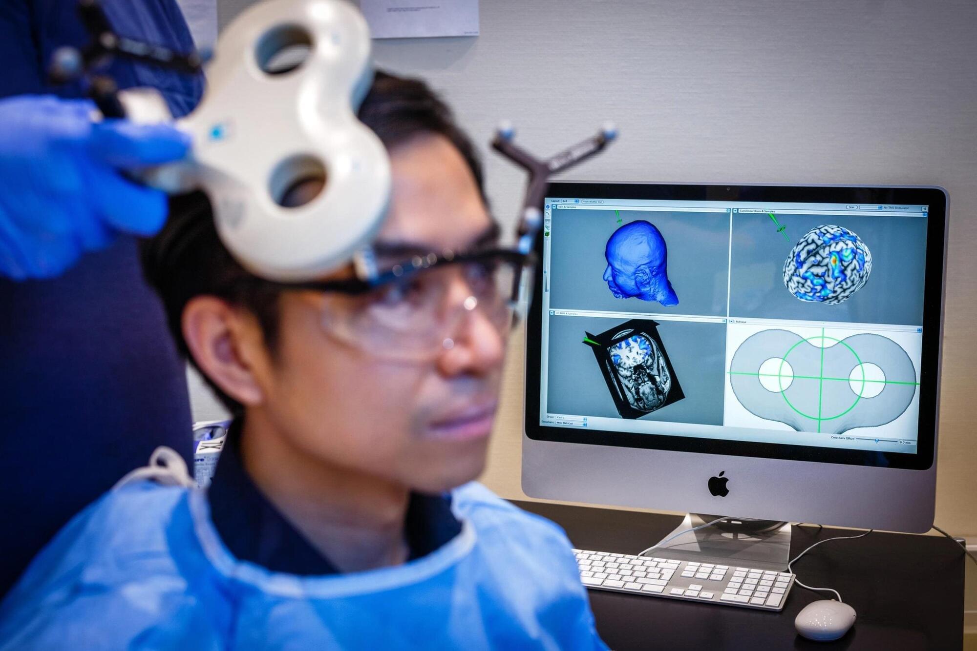
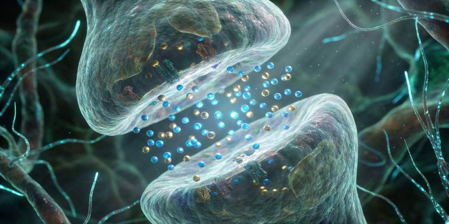
Scientists found that synchronizing activity between two brain regions made people more generous.
A new study suggests that synchronizing activity in specific parts of the brain can make people more likely to act generously. Research published today (February 10) in the open-access journal PLOS Biology reports that stimulating two brain regions in a coordinated way increased altruistic behavior. The study was led by Jie Hu of East China Normal University in China, working with colleagues from the University of Zurich in Switzerland.
Why some people are more altruistic than others.
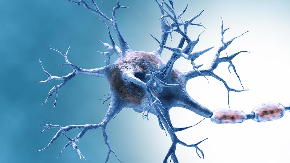
Our brains age along with the rest of our bodies, and as they do, they produce fewer new brain cells. Now, researchers have found a key mechanism through which the typical age-related decline in neuron production might be slowed.
In later life, the neural stem cells (NSCs) that turn into fully fledged neurons become more dormant – almost as if they’re going into retirement after a long lifetime of service. As that happens, cognitive decline creeps in.
A major reason why NSC activity fades with age is the wear and tear on telomeres, the protective caps on the ends of DNA. Telomeres fray a little more each time a cell divides, and over time, this impairs cells’ ability to grow and divide, leading to increasing cell death.
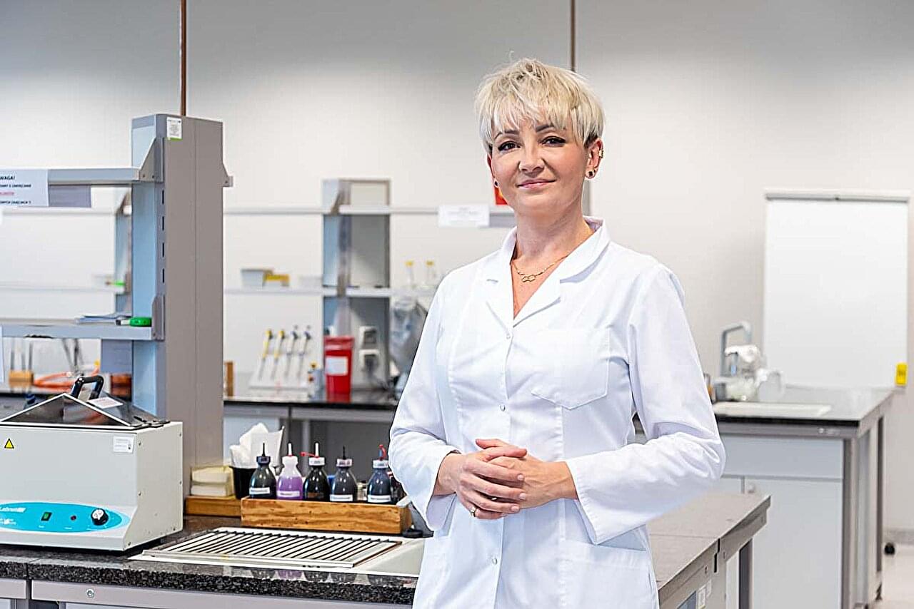
Miracle Mineral Solution, also known as MMS, has been marketed for years as a purported miracle cure for various conditions, including cancer, autism, and COVID-19. MMS is the marketing name for sodium chlorite (NaClO₂), a powerful disinfectant used, among other things, for water treatment. When sodium chlorite is acidified, chlorine dioxide (ClO₂) is formed. Its consumption can be hazardous to health.
A team of scientists from Wroclaw Medical University decided to investigate this.
In a study published in Scientific Reports, they analyzed the effects of acidified sodium chlorite (ASC), from which ClO₂ is produced.
This is a ~57 minute talk titled “The Bioelectric Interface to the Collective Intelligence of Morphogenesis: development, regeneration, cancer, and beyond” which I gave at a UCSF seminar for an audience of graduate students and post-docs in Biophysics, Bioinformatics, and Chemical Biology. I covered the role of bioelectricity as cognitive glue underlying high-level adaptive plasticity in living tissue, recent progress in exploiting that interface, and new developments in research platforms for this field.
This is an invited talk in BAMΞ’s Mathematical Phenomenology Sprint.
Cf. https://bamxi.org/research-activities/mathematical-phenomenology-sprint/
Organizing Institutions:
Bamberg Mathematical Consciousness Science Initiative (BAMΞ) https://bamxi.org.
& Association for Mathematical Consciousness Science (AMCS) https://amcs-community.org
Oxidative stress is a direct consequence of an excess in the body of so-called free radicals—reactive, unstable molecules that contain oxygen. Free radicals are normal metabolic by-products and also help to relay signals in the body. In turn, oxidative stress (an overload of these molecules) can be caused by lifestyle, environmental, and biological factors such as smoking, high alcohol consumption, poor diet, stress, pollution, radiation, industrial chemicals, and chronic inflammation.
When this occurs, it creates an imbalance between the production of free radicals and the body’s antioxidant defenses, which are responsible for neutralizing them.
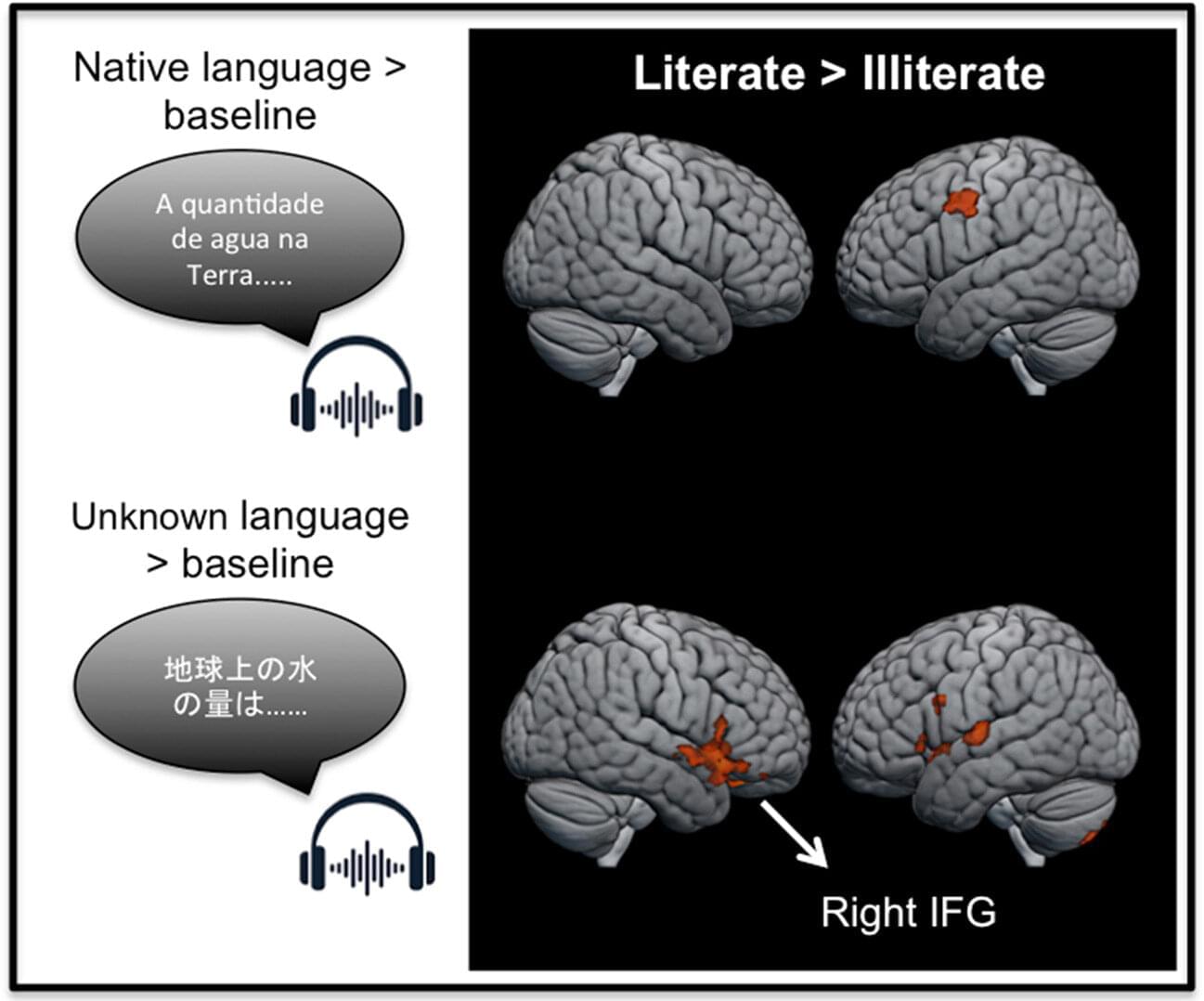
Learning to read reshapes how the brain processes language. New research from Baycrest and the University of São Paulo shows that learning to read fundamentally changes how the brain responds to spoken language, even when no written words are present. While previous brain imaging studies have demonstrated that literacy strongly affects how the brain responds to written words, this study is among the first to show differences in brain activity during listening alone.
The findings confirm that as people learn to read, they develop a skill known as phonemic awareness, the ability to hear and manipulate the individual sounds that make up spoken words, a core foundation of reading. The study shows that learning to read improves how the brain processes spoken language by increasing sensitivity to these component sounds. This, in turn, strengthens short-term verbal memory, supporting the ability to learn complex skills and manage the cognitive demands of daily life.
The work is published in the journal Cortex.
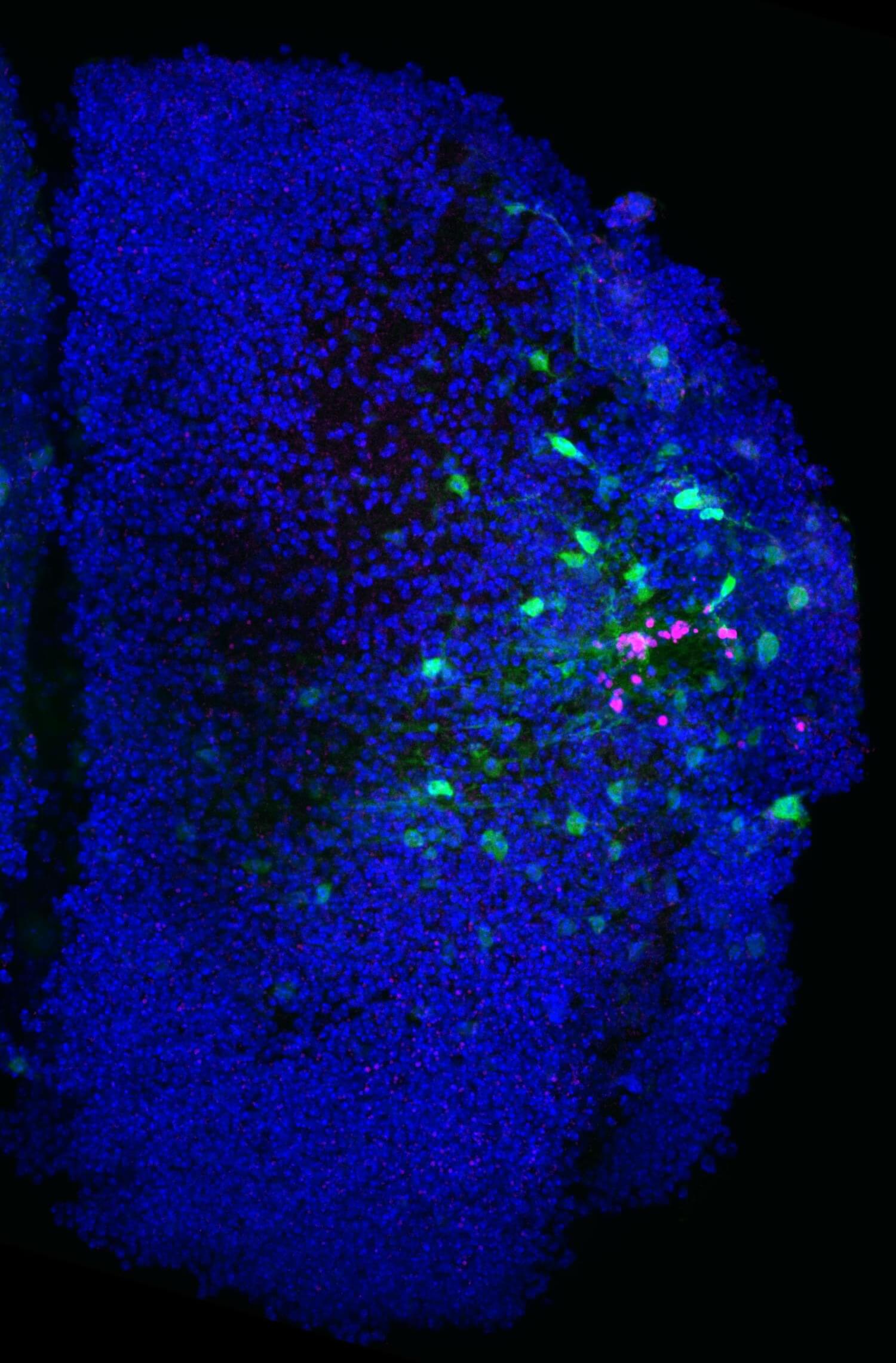
Could a hybrid biohardware using neural orgamoids and silicon make minduploading easier.
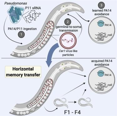
Animals face both external and internal dangers: pathogens threaten from the environment, and unstable genomic elements threaten from within. C. elegans protects itself from pathogens by “reading” bacterial small RNAs, using this information to both induce avoidance and transmit memories for four generations. Here, we found that memories can be transferred from either lysed animals or from conditioned media to naive animals via Cer1 retrotransposon-encoded virus-like particles. Moreover, Cer1 functions internally at the step of transmission of information from the germline to neurons and is required for learned avoidance. The presence of the Cer1 retrotransposon in wild C. elegans strains correlates with the ability to learn and inherit small-RNA-induced pathogen avoidance. Together, these results suggest that C.